About the Lab
Morphogenesis (= creation of a form; = shaping of an organism) is a fundamental aspect of developmental biology. Morphogenetic processes are based on self-organization, but, at the same time, are controlled by genetic regulatory networks and mechanical forces. At the tissue level, morphogenesis is based on cell shaping, cell proliferation/cell death, cell migration and cell adhesion. Evolutionary changes in morphogenetic processes are essential for the evolution of early development and the generation of evolutionary novelties including new body plans.
We explore the evolution of morphogenesis using the comparative approach. We study and compare cellular and molecular bases of morphogenesis in several model systems.
Research fields
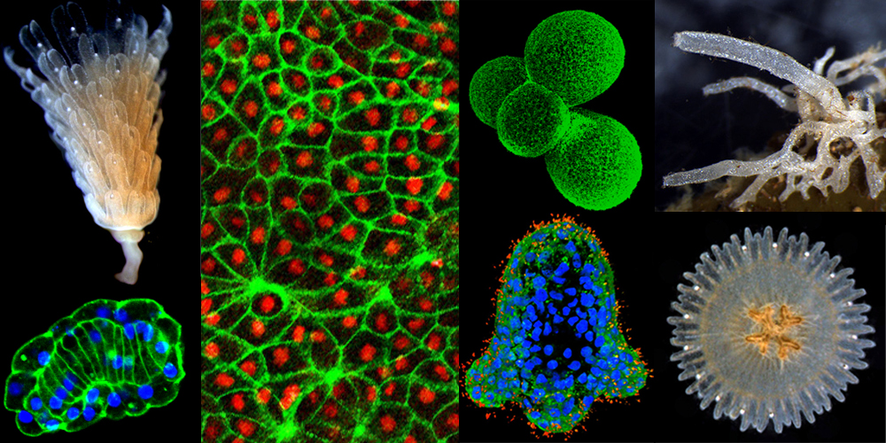
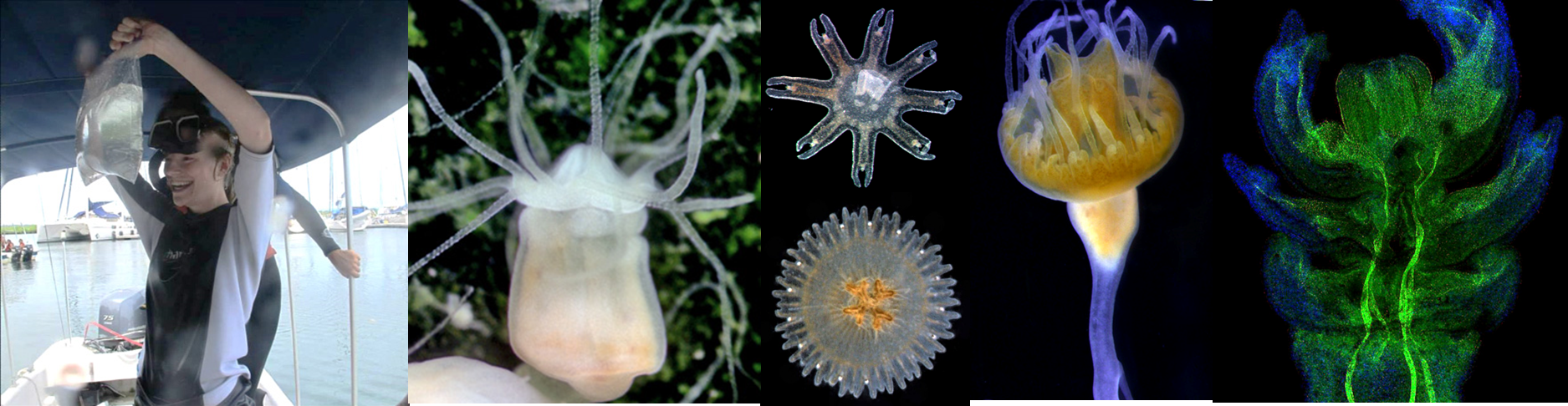
Evolution of Cnidarian's Early Development

Multicellular animals exhibit a fascinating diversity of morphologies. One of the outstanding questions in animal biology is the evolution of symmetry and polarity in body plans. To approach this question, we must improve our understanding of the patterning systems underlying the establishment of symmetry plans and body axes in the development of a wide range of animal species. Thus, we must have model objects, in which the general principles of patterning are tractable and accessible for experimental investigations.
Cnidaria as the sister group to all bilateral animals holds a key phylogenetic position to address early animal evolution. They are diploblastic animals composed of only two cell layers, ectoderm and endoderm. They are characterized by radial, tetrameric, and even bilateral symmetry. The embryonic and larval development is highly diverse in this group. Cnidarian body plans and developmental trajectories underwent multiple evolutionary modifications. This potentially provides an opportunity for comparative investigations within cnidarians and between Cnidaria and Bilateria. Unfortunately, the range of cnidarian model species does not cover the whole range of body plans and developmental histories evolved in this phylum, and our progress is strongly impeded by the lack of comparative data.
Our research team studies morphogenetic processes in the embryonic, larval and post-larval development of various cnidarian species: Dynamena pumila (Hydrozoa), Aglantha digitale (Hydrozoa), Aurelia aurita (Scyphozoa), Lucernaria quadricornis (Staurozoa) and others. Many of our species are not model objects and we collect embryos and larvae in natural populations. We use the methods of in vivo imaging, light microscopy, electron microscopy (SEM and TEM), confocal microscopy, histology, immunocytochemistry and molecular biology.
Evolution of Complex Life Cycle
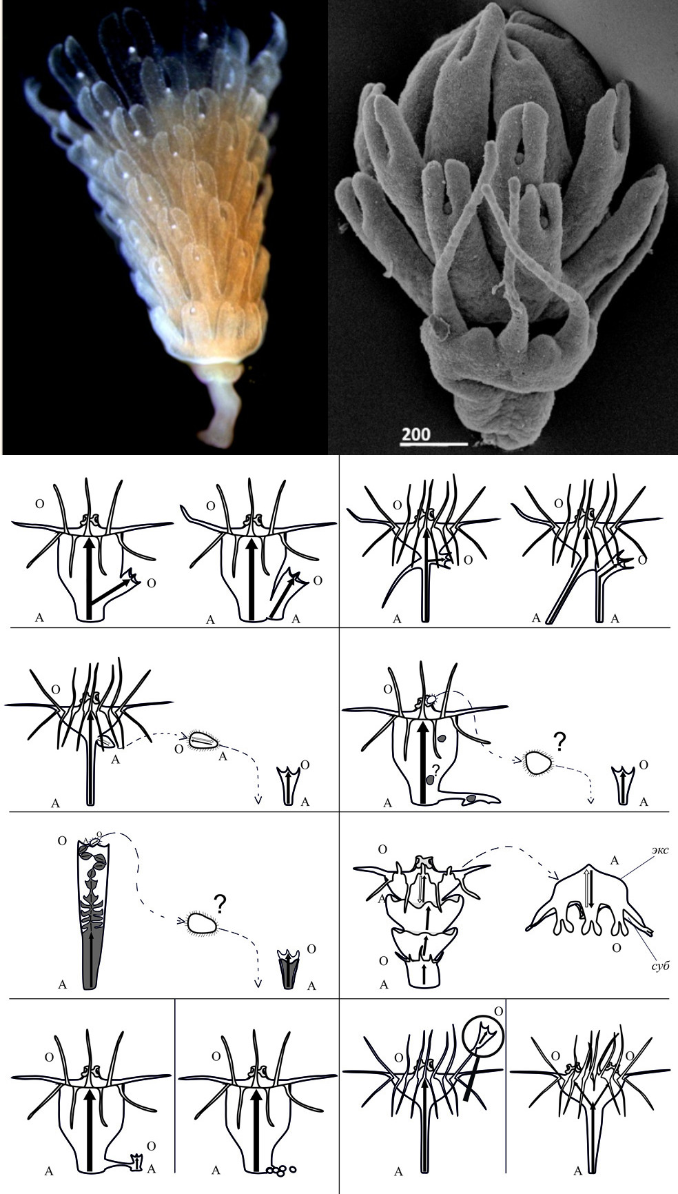
Fig. Modes of asexual reproduction in Scyphozoa (Sukhoputova et al., 2019)
The majority of metazoans possess complex life cycles, which incorporate ecologically discrete and morphologically distinct stages (Davidson et al., 1995; Raff, 2008). It seems that the origins or losses of complex life cycles are associated with the most important events in the formation of biodiversity. A number of hypotheses have been proposed to explain the origin and abundance of complex life cycles, but many fundamental questions concerning their evolution remain unanswered. First of all, it is unclear to what extent different stages are able to evolve independently of one another. In particular, if later developmental stages depend on earlier ones, how can an intermediate stage of development (e.g. the larval stage) be lost without deleterious effects? To address this question, we need to establish a model system, which should include several phylogenetically distant species exhibiting similar evolutionary modifications of their life cycles.
Holopelagic life cycle evolution
The Medusozoa is a group within the cnidarians comprising the classes Hydrozoa, Scyphozoa, Cubozoa and Staurozoa. The great advantage of this group is in the high level of evolutionary plasticity of their complex life cycle that has been subjected to multiple evolutionary modifications (Leclere et al., 2016). In the ‘typical’ medusozoan life cycle, a juvenile stage (polyp) asexually produces an adult stage (medusa); the medusa produces gametes which fuse and the resulting embryo develops into a planula larva; the planula metamorphoses into a sessile polyp. The evolutionary plasticity of the medusozoan life cycle is extremely high: there are many independent losses of the medusa/polyp stage within Hydrozoa and Scyphozoa. We will focus our studies on hydrozoan and scyphozoan species that have lost the polyp stage and acquired the so-called ‘holopelagic life cycle’.
Since the medusa is normally produced from polyp tissues by lateral budding (Hydrozoa) or by transverse fission (Scyphozoa), the loss of the polyp stage imposes severe developmental constraints. Holopelagic species had to substantially modify their embryonic and larval development to acquire the ability to form medusae directly from larvae, not from polyps. To gain insight into the rules of life cycle evolution, we need to investigate the developmental strategies allowing holopelagic medusae to overcome the constraints imposed by changes in the life cycle.
Acquiring the holopelagic life cycle is the main evolutionary trend in the hydrozoan subclass Trachylina, which includes some of the most interesting and enigmatic cnidarians. We study the embryonic and larval development of several Trachylina species: Aglantha digitale, Liriope tetraphilla, Solmundella bitentaculata.
Interestingly, the life cycle of the scyphozoan jellyfish Pelagia noctiluca underwent the same evolutionary modification. In Pelagia, the polyp stage has been lost and the larva directly transforms into an adult medusa in the course of metamorphosis.
Asexual reproduction in cnidarians
Scyphozoa is a large and diverse group of medusozoan cnidarians. Medusa formation in Scyphozoa occurs via strobilation, i.e. the fission of a polyp (scyphistoma) into future medusa discs by transverse constrictions. These discs undergo a series of developmental transformations and ephyra forms from each of them. The free-swimming ephyra detaches from the polyp and gradually transforms into a sexually mature medusa. A part of the polyp body (stalk) remains after the strobilation and transforms into the body of a new polyp. Strobilation can be considered the most common mode of asexual reproduction observed in scyphozoans. We study the morphogenetic processes underlying the strobilation in the scyphozoans Aurelia and Cassiopea.
Development and Regeneration of Sponges
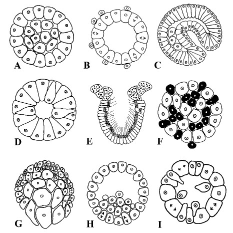
Fig. Different types of morphogenesis in Porifera resulting in larva formation (Ereskovsky, Dondua, 2006)
While virtually all animals show certain abilities for regeneration after an injury, these abilities vary greatly among metazoans. The reparative regeneration can occur at various scales: from the restoration of damaged cells, tissues and organs to the reconstruction of a whole body from a small portion of tissues, i.e., Whole-Body Regeneration (WBR). WBR can occur either with retaining the original organism’s polarity and symmetry or through a complete rebuilding of the tissues involved, accompanied by a de novo formation of body axes.
Porifera (Sponges) is a basal lineage of metazoans characterized by a particular body organization: they lack a gut, muscles, nerves and conventional neuronal signalling systems, organs and organ systems homologous to Eumetazoa. Sponges are capable of a wide variety of different regenerative processes: healing wounds, regenerating excised body parts, restoring from tissue fragments of various sizes and restoration during cell reaggregation.
The morphogenesis, cellular sources and dynamics of cell proliferation during reparative regeneration in sponges have been studied at histological and ultrastructural levels. These studies are consistent in denoting transdifferentiation as one of the essential mechanisms of reparative regeneration in sponges. However, the morphogenetic mechanisms underlying regeneration are fundamentally different in different clades of sponges. In sponges from the classes Homoscleromorpha and Calcarea, epithelial morphogenesis predominates during regeneration, and the primary cellular sources (pluripotent cells) for the restoration of the lost structures are choanocytes and pinacocytes. At the same time, Epithelial-to-Mesenchymal transformations (EMT) and mesenchymal morphogenesis dominate in the regeneration of Demospongiae, and archaeocytes and choanocytes are the primary cellular sources. Moreover, only in demosponges, regeneration is accompanied by the formation of a blastema, which is absent during regeneration in Homoscleromorpha and Calcarea.
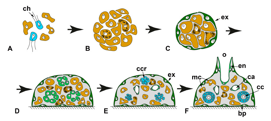
Fig. Whole Body Regeneration after tissue dissociation (cell reaggregation) (Ereskovsky et a., 2021)
However, it remains unclear if the molecular mechanisms governing WBR are conserved in phylogenetically distant sponge groups. This raises many questions: How conserved is the role of molecular mechanisms described for reparative regeneration? Are all molecular pathways involved in regeneration universal? Do taxon-restricted genes play key roles in regeneration processes? What molecules govern complex cellular behaviour (such as EMT) in the absence of key regulatory proteins that we know from high metazoans? All of these and many other questions outline the role of WBR in modern regenerative biology as a promising model for studying regeneration in invertebrates.
We currently work on the following projects.
- Cytoskeleton dynamic in sponges. Cell migration and reshaping play a leading role in embryonic development, regeneration, immune response etc. The spatiotemporal regulation of the cytoskeleton dynamics is one of the main mechanisms in these cell behaviours. Data on the cytoskeleton of the Porifera cells, one of the basal evolutionary branches of Metazoa, remain fragmentary. This project aims to obtain new data on the cytoskeleton in phylogenetically distant sponge species. Thanks to an integrative approach, we will obtain new data on the structure, dynamics, role and mutual regulation of cytoskeleton systems during the development and regeneration of sponges. These results will be very valuable for further research in the biology of both sponges and all Metazoa. The combination of classical and modern methods and approaches adopted in cell biology and developmental biology will allow for the first time to conduct a comprehensive study of the structure of the cytoskeleton of sponges from the classes Demospongiae and Calcarea, as well as the dynamics and role of the cytoskeleton in the morphogenesis accompanying the normal development and regeneration in these sponges.
- Investigation of the axialisation in sponges during whole-body regeneration. We hypothesized that during the development of cell aggregates, primmorphs, new axes are established due to the activation of the Wnt and TGF-beta signalling pathways. Using in silico analysis, RNA-seq and whole-mount in situ hybridization, we want to identify the participants in these signalling pathways and determine the spatiotemporal changes in their expression in demosponge Halisarca dujardinii and calcarean Leucosolenia variabilis.
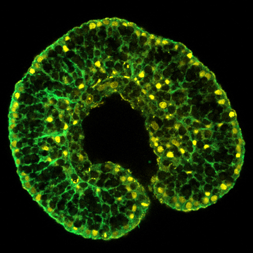
Comparative Embryology of Bilateria
Fig.
Fig.
The lab members

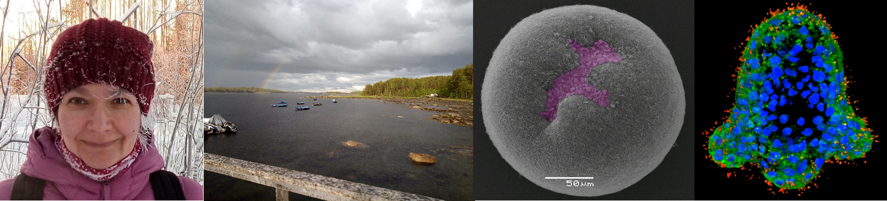
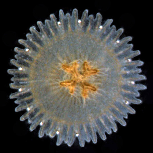
From left to right: Yulia Kraus; Our research site, White Sea Biological Station; Early gastrula of Nematostella vectensis (Anthozoa); Larva of Aglantha digitale (holopelagic hydromedusa)
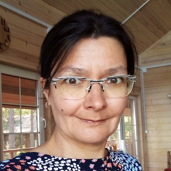
Yulia graduated from the Department of Evolutionary Biology (Faculty of Biology, Lomonosov Moscow State University). She defended her PhD thesis ‘Self-organization phenomena in the early development of marine hydroids’ in 1998.
"The main area of my research interest is the Evo-Devo of basal metazoans, in particular, the evolution of embryonic and larval development, organizers and axial patterning. The ongoing projects of my research team focus on the evolution of morphogenetic processes and complex life cycles in cnidarians. I have longstanding expertise (about 25 years) in research work on cnidarian embryos and larvae: I have already investigated the normal development of 11 cnidarian species and got skills in comparative embryology, experimental embryology, histology, electron microscopy, confocal microscopy, immunocytochemistry and molecular biology."
Yulia also works at the Department of Evolutionary Biology (MSU Faculty of Biology). Her teaching activity includes the following courses:
Evolutionary Developmental Biology (for Master students).
Analysis and Writing of Scientific Papers (for Bachelor students).
Ontogeny Evolution (for Bachelor students).
Evolutionary Theory (for Bachelor students).
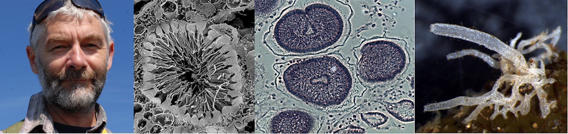
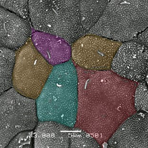
From left to right: Alexander Ereskovsky; Choanocyte chamber of Oscarella lobularis (Homoscleromorpha) (SEM); Developing embryos of Halisarca dujardinii (Demospongiae); Leucosolenia variabilis (Calcarea)
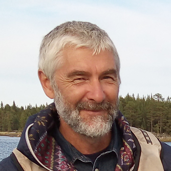
Alexander graduated from the Department of Embryology of the Saint-Petersburg State University (Russia).
Alexander studies the development of sponges from different clades. He performs comparative analyses of their development to gain insight into the evolution of sponge embryonic and postembryonic development and regeneration. His studies help to clarify the evolutionary origin of a variety of key morphogenetic mechanisms and developmental processes, including epithelial and mesenchymal morphogeneses of embryos and adults. Alexander is also interested in the evolution of life cycles, phenology and reproductive cycles of marine invertebrates, major transitions in animal evolution, such as the emergence of multicellularity and morphological complexity, and the evolution of ontogeny of basal metazoans.
Alexander conducts his research work focusing on embryology and evolutionary biology not only at the IDB RAS but also at the Saint-Petersburg State University (Russia) and at the Mediterranean Institute of Biodiversity and Ecology (CNRS, Aix-Marseille University, Marseille, France).
Stanislav (Stas) graduated from the Department of Embryology (Faculty of Biology, Moscow State University). In 2012, he defended his PhD thesis "Mechanodependent cellular and molecular processes in the epithelial morphogenesis of Xenopus laevis embryos". He works in the area of Evolutionary Developmental Biology (Evo-Devo). He is interested in the evolution of molecular mechanisms underlying body plan formation during the embryonic and larval development of metazoans.
"Molecular pre-patterning during animal development is a fundamental process that underlies all developmental events, including body plan establishment, cell differentiation, tissue and organ formation, and, finally, a proper functioning of all systems of the adult body. It is surprising that different animals with different body plans use the same molecular tool kits in the course of development. It seems that evolutionarily conserved signaling pathways should be fine-tuned to participate in the development of different body plans, tissues and organs. Which regulatory mechanisms provide this fine-tuning? To answer this question, I use different model animals, both vertebrates and invertebrates, to study various developmental processes including the establishment of the body plan and differentiation of the body axes."
Stanislav also works at the Department of Embryology (MSU). He gives lectures on the Molecular Methods of Developmental Biology.
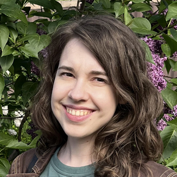
Alena graduated from the Faculty of Biology of Moscow State University in 2013. In 2019, she defended her PhD thesis "Comparative Analysis of Strobilation in Scyphozoan Representatives".
Her main area of interest is evolutionary developmental biology (Evo-Devo) and comparative morphology of basal metazoans. She has mastered in the methods of light and electron microscopy and immunocytochemistry. Alena studies the typical life cycle of scyphozoans that includes the planula, polyp, ephyra (larval medusa) and medusa stages. Strobilation is a major mode of asexual reproduction of the scyphozoan polyps. At the same time, strobilation provides the transition between the polyp and medusa life forms. Alena investigates the morphogenetic processes underlying strobilation. She characterizes cell shape changes, cell behavior and proliferation at the successive stages of the ephyra formation.
Alena teaches the following courses at the Department of Evolutionary Biology (MSU): Analysis and Writing of Scientific Papers (for Bachelor students), Evolutionary Theory (for Bachelor students).
Sasha graduated from the Department of Embryology (Faculty of Biology, Lomonosov Moscow State University) with a Master's degree in Embryology in 2018. She is interested in how different modes of development shape body plans in marine invertebrates. Sasha studies the embryonic development of a marine hydroid, Dynamena pumila. Her PhD project explores the cellular and molecular mechanisms of axis formation during embryogenesis in cnidarians with apolar gastrulation (the term "apolar gastrulation" means that endoderm formation is not confined to a single specific area of the embryo).

Ksenia is a Research Assistant, but, at the same time, she is a PhD student.
Kseniia graduated from the Department of Cell Biology (Faculty of Biology, Moscow State University) in 2022.
Kseniia's research focuses on reparative morphogenesis in basal Metazoa (in the representatives of Porifera, in particular) and its underlying mechanisms. More specifically, she is interested in cytoskeletal rearrangements involved in the morphallactic and epimorphic pathways of regeneration and the signaling pathways that control them. She studies the phenomenon of actomyosin contractility during wound healing in sponges and its potential intracellular regulators. Kseniia employs a number of modern molecular techniques and a wide range of microscopic approaches.
The key step of regeneration in multicellular animals is epithelialization, since restoration of epithelial integrity is crucial. Two mechanisms are involved in epithelial restoration: the actomyosin cable "purse string" contraction and collective cell migration (Martin and Lewis, 1992; Zahm et al., 1991). However, the interaction between these two epithelialization pathways remains unclear, especially in basal metazoans (sponges, cnidarians, placozoans and ctenophores). It is known that epithelial tissues are the most ancient. Therefore, characterization of the mechanisms underlying their homeostasis and physiological/reparative renewal in extant animals may provide some clues about the evolution of multicellularity. These data can be obtained using an integrative approach combining classical morphology methods with modern molecular, genomic and transcriptomic techniques.
Grants & Publications
Grants
“The molecular mechanisms behind the life cycle evolution and speciation in hydroids of the Arctic region" - Russian Science Foundation (RSCF) Grant; PI - Stanislav Kremnyov.
Selected Publications
- Kraus Y., Osadchenko B, Kosevich I. Embryonic development of the moon jellyfish Aurelia aurita (Cnidaria, Scyphozoa): another variant on the theme of invagination//PeerJ. 2022. Vol. 10. Art no e13361. DOI: 10.7717/peerj.13361.
- Kremnev S.V. Evolutionary and Ontogenetic Plasticity of Conserved Signaling Pathways in Animals’ Development//Russian Journal of Developmental Biology. 2022. Vol. 53. № 2. P. 65–81. DOI: 10.1134/S1062360422020114.
- Petri N, Nordbrink R, Tsikolia N, Kremnyov S. Abnormal left-right organizer and laterality defects in Xenopus embryos after formin inhibitor SMIFH2 treatment // PLoS ONE. 2022. Vol.17. №11. e0275164. doi.org/10.1371/journal.pone.0275164
- Negretti M.I., Böse N., Petri N., Kremnyov S., Tsikolia N. Nodal asymmetry and hedgehog signaling during vertebrate left–right symmetry breaking // Frontiers in Cell and Developmental Biology. 2022. Vol.10. Art no. 957211. DOI: 10.3389/fcell.2022.957211
- Ereskovsky A.V., Tokina D. Ultrastructural research of spermiogenesis in two sponges, Crellomima imparidens and Hymedesmia irregularis (Demospongiae): New evidence of sperms with acrosome in sponges//Journal of Morphology. 2022. Vol. 283. №3. P. 333-345. DOI: 10.1002/jmor.21446.
- Lavrov A.I., Bolshakov F.V., Tokina D.B., Ereskovsky A.V. 2021. Fine details of the choanocyte filter apparatus in asconoid calcareous sponges (Porifera: Calcarea) revealed by ruthenium red fixation // Zoology. 2022. Vol. 150. Art.no. 125984. DOI: 10.1016/j.zool.2021.125984.
- Martinez P., Ballarin L., Ereskovsky A.V.3, Gazave E., Hobmayer B., Manni L.C., Rottinger E., Sprecher S.G., Tiozzo S.l., Varela-Coelho A., Rinkevich B. Articulating the "stem cell niche" paradigm through the lens of non-model aquatic invertebrates / /BMC Biology. 2022. Vol. 20. №. 1. Art.no. 23. DOI: 10.1186/s12915-022-01230-5.
- Ereskovsky A., Borisenko I.E, Bolshakov F.V., Lavrov A.I. 2021. Whole-body regeneration in sponges: diversity, fine mechanisms and future prospects. Genes. 12, 506. https://doi.org/10.3390/genes12040506
- Ereskovsky, A., Lavrov, A. 2021. Porifera. In Invertebrate Histology, Elise E.B. LaDouceur, E.E.B., Ed., John Wiley & Sons, Inc. https://doi.org/10.1002/9781119507697.ch2
- Mayorova T.D., Osadchenko B., Kraus Y. How to build a larval body with less than a hundred cells? Insights from the early development of a stalked jellyfish (Staurozoa, Cnidaria)//Organisms Diversity & Evolution. – 2020. DOI: 10.1007/s13127-020-00459-8. – Q1.
- Kraus Y., Chevalier S., Houliston E. Cell shape changes during larval body plan development in Clytia hemisphaerica // Developmental Biology. – 2020. DOI: 10.1101/864223. (WoS, Scopus) – Q2.
- Kupaeva D., Konorov E., Kremnyov S. De novo transcriptome sequencing of the thecate colonial hydrozoan, Dynamena pumila//Marine Genomics. – 2020. DOI: https://doi.org/10.1016/j.margen.2019.100726. (WoS, Scopus) – Q3.
van der Sande M., Kraus Y., Houliston E., Kaandorp J. A cell-based boundary model of gastrulation by unipolar ingression in the hydrozoan cnidarian Clytia hemisphaerica // Developmental Biology. 2020. DOI: 10.1016/j.ydbio.2019.12.012. (WoS, Scopus) – Q2. - Ereskovsky A.V., Tokina D. B., Baghdiguian S., Le Goff E., Lavrov A. I. 2020. Transdifferentiation and mesenchymal-to-epithelial transition during regeneration in Demospongiae (Porifera). Journal of Experimental Zoology Part B: Molecular and Developmental Evolution. 334: 37-58. DOI: 10.1002/jez.b.22919
- Ereskovsky A.V. 2019. In search of ancestral organization of sponges Bauplan and phylotypic stage in Porifera. Russian Journal of Developmental biology. 50(6): 398–406. DOI: 10.1134/S1062360419060031
- Nikishin D.A., Rimskaya-Korsakova N.N., Kremnyov S.V., Bagaeva T.S., Khramova Y.V., Semenova M.L., Kosevich I.A., Kraus Y.A., Vortsepneva E.V., Lavrov A.I., Prudkovsky A.A. Atlas of the White Sea Invertebrates Development. 3rd Summer Course on Embryology of Marine Invertebrates, June 9-30, 2019, WSBS MSU, Russia. — М.Pero, 2019. — 44 с. ISBN 978-500150-258-6.
- Bagaeva T.S., Kupaeva D.M., Vetrova A.A., (...), Kraus Y.A., Kremnyov S.V. cWnt signaling modulation results in a change of the colony architecture in a hydrozoan // Developmental Biology. 2019. DOI: 10.1016/j.ydbio.2019.08.019. (WoS, Scopus) – Q2.
- Kupaeva D.M., Vetrova A.A., Kraus Y.A., Kremnyov S.V. Epithelial folding in the morphogenesis of the colonial marine hydrozoan, Dynamena pumila // Biosystems. 2018. V. 173. P. 157-164. DOI: 10.1016/j.biosystems.2018.09.008. (WoS, Scopus).
- Kirillova A., Genikhovich G., Demilly A., Pukhlyakova E., Kraus Y., Technau U. Germ layer commitment and axis formation in sea anemone embryonic cell aggregates// Proceedings of the National Academy of Sciences. 2018. V. 115. № 8. P. 1813–1818.
- Kremnyov, S., Henningfeld, K., Viebahn, C., Tsikolia, N. Divergent axial morphogenesis and early shh expression in vertebrate prospective floor plate // EvoDevo. 2018. V. 9. (1). P. 4.
Library & Useful Links
Library
Under construction
Useful Links
Nikolai Pertsov White Sea Biological Station (WSBS) of Lomonosov Moscow State University (MSU), http://en.wsbs-msu.ru/ - WSBS MSU is an educational and research center, created for conducting marine scientific research and student field practical courses at the White Sea. WSBS is a subdivision of the MSU Faculty of Biology. WSBS MSU is the base for the Research & Education Center "Marine Biology, Oceanography and Geology".
European Society for Evolutionary Developmental Biology (Euro Evo Devo) https://evodevo.eu/
The aim of the society is to promote evolutionary developmental biology by regularly organizing meetings on this subject in Europe.
The Biodiversity Heritage Library https://www.biodiversitylibrary.org/
The Biodiversity Heritage Library improves research methodology by collaboratively making biodiversity literature openly available to the world as part of a global biodiversity community.
iBiology https://www.ibiology.org/
iBiology’s mission is to convey, in the form of open-access free videos, the excitement of modern biology and the process by which scientific discoveries are made.
News & Events
Under construction