About the Research group
Embryophysiology is a subunit of Regenerative Biology Lab.
Classic neurotransmitters – serotonin, catecholamines, acetylcholine, etc. appear in the ontogenesis far before first neurons and perform many regulatory and modulatory functions. The induction of oocyte maturation, control of cell cycle during cleavage divisions, regulation of ciliary motility, the establishment of right-left asymmetry, and regulation of morphogenetic processes are part of the list of the transmitter’s embryonic functions. It is noteworthy that the prenervous activity of neurotransmitters has been described in representatives of various groups of the animal kingdom, including sponges and even protozoa. The Group of Embryophysiology is studying mechanisms of transmitter regulation of early embryonic development and oogenesis.
The research of the Group has been carried out in the frame of Section “Transmitter mechanisms of gametogenesis and early embryogenesis” of IDB RAS Government basic research program in 2021 № GZ 0088-2021-0009 “Mechanisms of the ontogenesis regulation: gametogenesis, fertilization, and early animal development.”
Group Members
Denis Nikishin
Head of the group
Senior Researcher, PhD
Field of interests: molecular mechanisms of transmitter regulations of the early embryogenesis and oogenesis.
Yulia Nikishina
Researcher, PhD
Field of interests: the role of catecholamines in development and ovarian function.
Nina Alyoshina
Researcher, PhD
Field of interests: neurotransmitter regulation of oogenesis and folliculogenesis.
Lyudmila Mal’chenko
Researcher-Engineer
Specialist in cell culture, immunohistochemical, and radioisotope analysis.
Veronica Frolova
PhD Student
Thesis research is devoted to studying the structure-functional organization of the serotonergic system in the early embryo cells.

Maria Tkachenko
PhD candidate
Thesis research is devoted to studying molecular mechanisms of the negative effects of serotonin uptake inhibition on oocyte maturation and early development.
Directions of Research
Structural and functional organization of transmitter signaling systems in the cells of the early embryo
Complex study of the expression of the receptors, transporters, and other components of transmitter systems, as well as their intracellular distribution, including co-localization with intracellular structures, is carried out in the early embryos of mammals, sea urchins, and clawed frogs.
It was shown that the embryonic mechanisms of transmitter systems are based on the same critical components as in adult cells, including synaptic structures. At the same time, embryonic mechanisms have some peculiarities. First, several transmitters and the corresponding number of receptors may be present and functionally active inside the embryonic cell. Second, the signal function of the transmitter, including its binding to the receptor or its functional analog, at least in some cases, happens on the intracellular level during cleavage divisions. It is the principal difference of embryonic cells from the differentiated ones, although this rule has exclusions. A fundamentally new question that has not been raised before in world literature is the possible simultaneous presence and functional activity of several receptor proteins activated by one transmitter in the same embryonic cell.
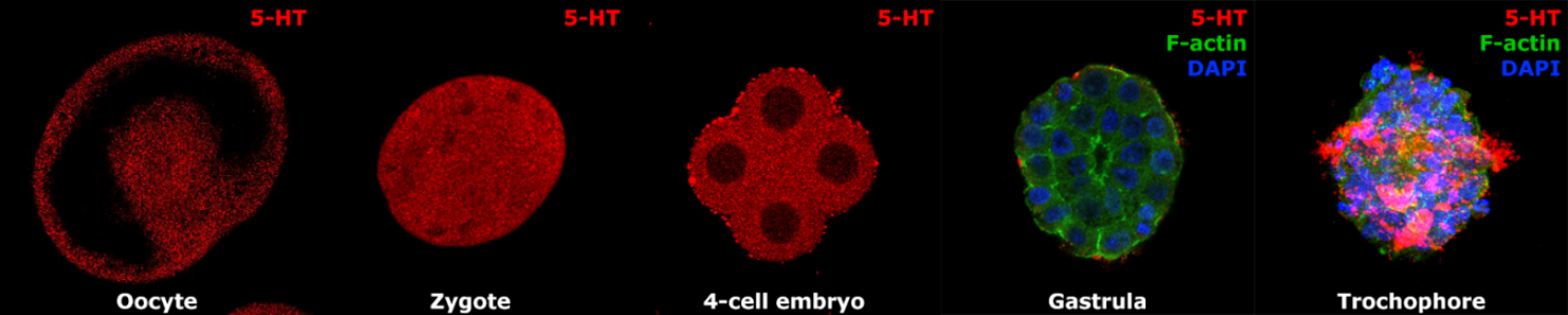
Fig. 1 Serotonin immunoreactivity in the developing embryos and larvae of polychaete Ophelia limacine. Serotonin is present in the cytoplasm of oocytes and early embryos during cleavage divisions but almost disappears at the gastrula stage. Later in development, it is detected mainly in neurons and in the area of the ciliary band.
Selected publications:
- Frolova, V. S., Nikishina, Y. O., Shmukler, Y. B., and Nikishin, D. A. Serotonin signaling in mouse preimplantation development: Insights from transcriptomic and structural-functional analyses. International Journal of Molecular Sciences 25, 23 (2024), 12954. DOI: 10.3390/ijms252312954
- Frolova, V. S., Ivanova, A. D., Konorova, M. S., Shmukler, Y. B., and Nikishin, D. A. Spatial organization of the components of the serotonergic system in the early mouse development. Biochemistry (Moscow), Supplement Series A: Membrane and Cell Biology 17, 1 (2023), 59–64. DOI: 10.1134/S1990747823060041
- Shmukler Y.B., Nikishin D.A., Non-Neuronal Transmitter Systems in Bacteria, Non-Nervous Eukaryotes, and Invertebrate Embryos // Biomolecules. 2022. V. 12, № 2. - P. 271. DOI: 10.3390/biom12020271
- Nikishin D.A., Khramova Y.V., Alyoshina N.M., Malchenko L.A., Shmukler Y.B. Oocyte-mediated effect of serotonin on the functional status of granulosa cells // Russian Journal of Developmental Biology. 2021. V. 52., № 2. - P. 97-104. DOI: 10.1134/S1062360421020065
- Nikishin D.A., Khramova Yu V., Bagayeva T.S., Semenova M.L., Shmukler Yu B. Expression of components of the serotonergic system in folliculogenesis and preimplantation development in mice // Russian Journal of Developmental Biology. 2018. V. 49., № 3. P. 184-192. DOI: 10.1134/S1062360418030062
- Shmukler Yu. B. and Nikishin D. A. On the Intracellular Transmitter Reception// Neurochemical Journal. 2018. Vol. 12, No. 4, P. 295–298 (2018). DOI: 10.1134/S1819712418040074
- Shmukler Yu. B. and Nikishin D. A. Transmitter systems in embryogenesis – current state of the problem // Uspekhi Fiziol. Nauk. 2018. V. 49.,. № 4. - P. 81–92 (in Russian) DOI: 10.7868/S0301179818040069
- Nikishin D.A., Khramova Y. V., Kremnyov S. V., Shmukler Y.B. Conservativeness and features of pre-nervous serotonergic signaling system in early embryonic development // FEBS J. 2014. V. 281.,. № Special Issue: 40th FEBS Congress, The Biochemical Basis of Life. - P. 65–784.
- Nikishin D. A., Semenova M. N., Yu. B. Shmukler. Expression of transmitter receptor genes in early development of sea urchin Paracentrotus lividus // Russ.J. Developm. Biol., 2012. V. 43.,. № 3. - P. 212–216. DOI: 10.1134/S1062360412030058
- Shmukler Y.B., Nikishin D.A. Transmitters in Blastomere Interactions // Cell Interaction. : InTech, 2012. P. 31–66.
- Nikishin D.A., Kremnyov S.V, Konduktorova V.V, Shmukler Y.B. Expression of serotonergic system components during early Xenopus embryogenesis // Int. J. Dev. Biol. 2012. V. 56.,. № 5. - P. 385–391. DOI: 10.1387/ijdb.113475dn
Mechanisms of the action of transmitters and their functional analogs in the early embryonic development
These studies are carried out in the embryos of sea urchins, a classic model of blockade of the first cleavage division that allows to evaluate the effects of embryotoxic effects quantitatively. The influence on the cytoskeleton elements mediates cytotoxic effects of the antagonists of serotonin and dopamine receptors. They both evoke an increase in actin polymerization and disturbance of the cleavage spindle.
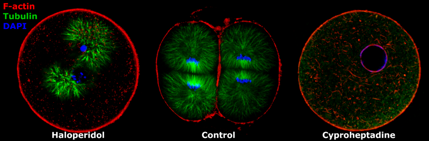
Fig. 2 Blockage of first cleavage division of sea urchin Paracentrotus lividus by the dopamine receptor antagonist haloperidol (left) and serotonin receptor antagonist cyproheptadine (right). In control - the normally cleaved 2-cell embryo. (Nikishin et al., 2020)
Selected publications:
- Nikishin D.A., Malchenko L.A., Milošević I., Rakić L., and Shmukler Y.B. Effects of Haloperidol and Cyproheptadine on the Cytoskeleton of the Sea Urchin Embryos // Biochemistry (Moscow), Supplement Series A: Membrane and Cell Biology, 2020, Vol. 14, No. 3, pp. 249–254. DOI: 10.1134/s1990747820020087
- Nikishin D.A., Milošević I., Gojković M., Rakić L., Bezuglov V.V, Shmukler Y.B. Expression and functional activity of neurotransmitter system components in sea urchins’ early development. // Zygote. 2016. V. 24.,. № 2. - P. 206–18. DOi: 10.1017/S0967199415000040
The role of serotonin in the regulation of mammalian oogenesis and ovarian folliculogenesis
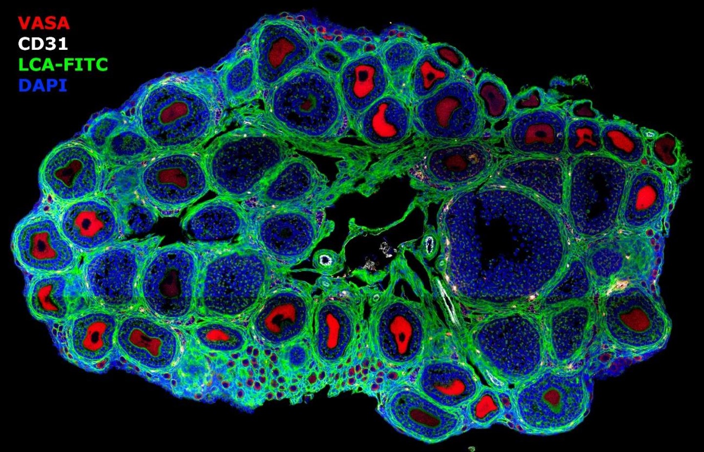
Fig. 3 Ovary of the prepubertal mouse during the first wave of folliculogenesis (10-17 dpp) is a useful experimental model that allows studying the state of ovarian pool and gonadotrope-independent period of early folliculogenesis.
Serotonin is one of the oocyte maturation factors in a lot of species. Among others, serotonin stimulates folliculogenesis in the mammalian ovary. It was shown that the functional activity of monoaminergic transmitters appears in oogenesis when oocyte maturation occurs and the base for further embryonic development forms. The main mechanism that determines the functional activity of serotonin in the mammalian ovary is its uptake by oocytes of growing ovarian follicles. The study of serotonergic regulation of folliculogenesis and female reproductive function, including side effects of antidepressants, first of all – selective inhibitors of serotonin re-uptake, are in progress in the Group.
Selected publications:
- Alyoshina Nina M., Tkachenko Maria D., Nikishina Yulia O., Nikishin Denis A. Serotonin Transporter Activity in Mouse Oocytes Is a Positive Indicator of Follicular Growth and Oocyte Maturity // Int. J. Mol. Sci. 2023. V. 24., № 14. P. 11247. DOI: 10.3390/ijms241411247
- Alyoshina Nina, Tkachenko Maria, Malchenko Lyudmila, Shmukler Yuri, Nikishin Denis A. Uptake and Metabolization of Serotonin by Granulosa Cells Form a Functional Barrier in the Mouse Ovary // Int. J. Mol. Sci. 2022. V. 23., № 23. P. 14828. DOI: 10.3390/ijms232314828
- Nikishin D.A., Khramova Y.V., Alyoshina N.M., Mal’chenko L.A., Shmukler Y.B. Oocyte-Mediated Effect of Serotonin on the Functional Status of Granulosa Cells // Russ. J.Dev.Biol. 2021. V. 52.,. № 2. - P. 97 - 104. DOI: 10.1134/S1062360421020065
- Alyoshina N.M., Malchenko L.A., Shmukler Y.B., Nikishin D.A. Specific serotonin uptake as a functional indicator of the developmental potential of follicles in the mouse ovary // FEBS open bio. 2021. V. 11., № S1. - P. P–03.1–24. DOI: 10.1002/2211-5463.13205
- Shmukler Y.B., Alyoshina N.M., Malchenko L.A., Nikishin D.A. Serotonin System in Oogenesis of Mammals // Neuroscience and Behavioral Physiology. 2022. V. 52., - P. 52–61. DOI: 10.1007/s11055-022-01207-5
- Nikishin D.A., Alyoshina N.M., Semenova M.L., Shmukler Y.B. Analysis of expression and functional activity of aromatic L-amino acid decarboxylase (DDC) and serotonin transporter (SERT) as potential sources of serotonin in mouse ovary // Int. J. Mol. Sci. 2019. V. 20.,. № 12. - P. 3070. DOI: 10.3390/ijms20123070
- Nikishin D., Alyoshina N., Shmukler Y. Expression and functional activity of specific membrane transport of serotonin in the mouse ovary // FEBS Open Bio. 2018. V. 8.,. № S1. - P. 381. DOI: 10.1002/2211-5463
- Nikishin, D.A., Alyoshina N.M., Shmukler Y.B. Synthesis and membrane transport of serotonin in the developing ovarian follicle of mouse // Doklady Biochemistry and Biophysics. 2018. V. 478., № 1. P. 4-7. DOI: 10.1134/s1607672918010027
- Nikishin D.A., Alyoshina N.M., Semenova M.L., Shmukler Y.B. Expression dynamics of the serotonergic system components in granulosa cells of the developing ovarian follicle and after luteinization // Genes and Cells. 2017. V. XII.,. № 4. - P. 33–38. (in Russian)
Financial Support
Grants of Russian Scientific Fundation (RSF)
- RSF 18-74-00143 – The Effect of Antidepressants of a Group of Selective Serotonin Reuptake Inhibitors (SSRIs) on Female Reproductive Function
- RSF 23-24-00621 – The Role of L-DOPA in the Pathogenesis of Polycystic Ovarian Syndrome and it's Treatment.
Grants of Russian Fund for Fundamental Researches (RFFI)
- RFBR 20-04-00303 – Structural and functional organization of monoaminergic transmitter signaling systems in oogenesis and early development of mammals
- RFBR 16-34-60250 – Study of molecular mechanisms of the serotonin regulation of proliferation, differentiation, and functional activity of granulosa cells
- RFBR 16-34-01217 – Role of serotonergic signal system in the regulation of the processes of growth and maturation of mammalian ovarian follicles
Grant of the President of the Russian Federation
- President Grant MK-931.2020.4 – Structural and functional organization of serotonergic signal system in oogenesis and early embryonic development
- President Grant MK-1304.2017.4 – Serotonin as local regulator of growth and development of mammalian ovarian follicles
Collaborations
- Department of Embryology of Biological Faculty, M.V. Lomonosov Moscow State university
- Lab of oxylipines of M.M. Shemyakin and Y.A. Ovchinnikov Institute of bioorganic Chemistry RAS
- Serbian Academy of Science and art (Belgrade, Serbia). Joint SASA and RAS «Neurotransmitters – Ontogenetic and Neurobiological Aspects»
- Institute of marine biology (Dobrota, Montenegro).
- Zoological Station “Anton Dohrn” (Naples, Italy). Grant ASSEMBLE Plus №8460 – Transmitters in the control of sea urchin embryo cytoskeleton
Selected publications of the Research Group
- Nikishin D.A., Khramova Y.V., Alyoshina N.M., Malchenko L.A., Shmukler Y.B. Oocyte-mediated effect of serotonin on the functional status of granulose cells // Russian Journal of Developmental Biology. 2021. V. 52., № 2. - P. 97-104.
- Shmukler Y.B., Alyoshina N.M., Malchenko L.A., Nikishin D.A. Serotonin System in Oogenesis of Mammals // Neuroscience and Behavioral Physiology. 2021. V. 71.,. № 3. - P. 306–320.
- Alyoshina N.M., Malchenko L.A., Shmukler Y.B., Nikishin D.A. Specific serotonin uptake as a functional indicator of the developmental potential of follicles in the mouse ovary // FEBS open bio. 2021. V. 11., № S1. - P. P–03.1–24.
- Nikishin D.A., Malchenko L.A., Milošević I., Rakić L., Shmukler Y.B. Effects of Haloperidol and Cyproheptadine on the Cytoskeleton of the Sea Urchin Embryos // Biochem. Moscow Suppl. Ser. A: Membrane and Cell Biology. 2020. № 14. - P. 249-254.
- Filatov M.A., Nikishin D.A., Khramova Y.V., Semenova M.L. Reference genes selection for real-time quantitative PCR analysis in mouse germinal vesicle oocytes // Zygote. 2019. № 2007. - P. 1–6.
- Nikishin D.A., Alyoshina N.M., Semenova M.L., Shmukler Y.B. Analysis of expression and functional activity of aromatic L-amino acid decarboxylase (DDC) and serotonin transporter (SERT) as potential sources of serotonin in mouse ovary // Int. J. Mol. Sci. 2019. V. 20.,. № 12. - P. 3070.
- Gorkun A.A., Shpichka A.I., Zurina I.M., Koroleva A. V, Kosheleva N. V, Nikishin D.A., Butnaru D. V, Timashev P.S., Repin V.S., Saburina I.N. Angiogenic potential of spheroids from umbilical cord and adipose-derived multipotent mesenchymal stromal cells within fibrin gel // Biomed. Mater. 2018. V. 13.,. № 4. - P. 044108.
- Nikishin D.A., Filatov M.A., Kiseleva M. V, Bagaeva T.S., Konduktorova V. V, Khramova Y. V, Malinova I. V, Komarova E. V, Semenova M.L. Selection of stable expressed reference genes in native and vitrified/thawed human ovarian tissue for analysis by qRT-PCR and Western blot // J. Assist. Reprod. Genet. 2018. V. 35.,. № 10. - P. 1851–1860.
- Nikishin D.A., Khramova Yu V., Bagayeva T.S., Semenova M.L., Shmukler Yu B. Expression of components of the serotonergic system in folliculogenesis and preimplantation development in mice // Russian Journal of Developmental Biology. 2018. V. 49., № 3. P. 184-192.
- Nikishin D., Alyoshina N., Shmukler Y. Expression and functional activity of specific membrane transport of serotonin in the mouse ovary // FEBS Open Bio. 2018. V. 8.,. № S1. - P. 381.
- Nikishin, D.A., Alyoshina N.M., Shmukler Y.B. Synthesis and membrane transport of serotonin in the developing ovarian follicle of mouse // Doklady Biochemistry and Biophysics. V. 478., № 1. P. 4-7.
- Vasilegina Y.I., Kremnev S. V., Nikishin D.A. Effects of mechanical stretching of embryonic tissues on axial structure formation in Xenopus laevis // Russ. J. Dev. Biol. 2017. V. 48.,. № 1. - P. 34–40.
- Nikishin D., Shmukler Y., Khramova Y. Serotonergic signaling system in granulosa cells of the developing ovarian follicle // FEBS J. 2017. V. 284.,. - P. 102–403.
- Shmukler Y.B., Nikishin D.A. Ladder-shaped ion channel ligands: current state of knowledge. // Mar. Drugs. 2017. V. 15.,. № 7.
- Nikishin D.A., Alyoshina N.M., Semenova M.L., Shmukler Y.B. Expression dynamics of the serotonergic system components in granulosa cells of the developing ovarian follicle and after luteinization // Genes and Cells. 2017. V. XII.,. № 4. - P. 33–38.
- Bondarenko N.S., Kurenkova A.D., Nikishin D.A., Umarova B.A. Effect of prolyl-glycyl-proline (PGP) and its acetylated form (N-AcPGP) on calcium level in the cytoplasm of rat peritoneal mast cells // Bull. Exp. Biol. Med. 2016. V. 161.,. № 4. - P. 487–490.
- Nikishin D.A., Milošević I., Gojković M., Rakić L., Bezuglov V. V, Shmukler Y.B. Expression and functional activity of neurotransmitter system components in sea urchins’ early development. // Zygote. 2016. V. 24.,. № 2. - P. 206–18.
- Brodsky V.Y., Malchenko L.A., Butorina N.N., Konchenko D.S., Zvezdina N.D., Dubovaya T.K. Glutamic Acid as Enhancer of Protein Synthesis Kinetics in Hepatocytes from Old Rats // Biochemistry (Mosc). 2017. V. 82., № 8. P. 957-961.
- Silina S.G., Nikishin D.A., Kremnyov S.V. Spatio-temporal expression pattern of mechanosensitive TRP ion channels during early development of Xenopus tropicalis // Biochemistry (Moscow) Supplement Series A: Membrane and Cell Biology. 2015. V. 9., № 3. P. 194-201.
- Nikishin D.A., Kremnyov S.V., Glagoleva N.S. Role of gap junctions and mechanosensitive ion channels in the mechanisms of growth pulsations of Gonothyraea loveni // Doklady Biological Sciences. 2015. V. 460., № 1. P. 64-67.
- Nikishin D.A., Khramova Y. V., Kremnyov S. V., Shmukler Y.B. Conservativeness and features of pre-nervous serotonergic signaling system in early embryonic development // FEBS J. 2014. V. 281.,. № Special Issue: 40th FEBS Congress, The Biochemical Basis of Life. - P. 65–784.
- Nikishin D.A., Semenova M.N., Shmukler Y.B. Expression of transmitter receptor genes in early development of sea urchin Paracentrotus lividus // Russ J Dev Biol. 2012. V. 43., № 3. P. 181-184.
- Shmukler Y.B., Nikishin D.A. Transmitters in Blastomere Interactions // Cell Interaction. : InTech, 2012. P. 31–66.
- Nikishin D.A., Kremnyov S.V, Konduktorova V.V, Shmukler Y.B. Expression of serotonergic system components during early Xenopus embryogenesis // Int. J. Dev. Biol. 2012. V. 56.,. № 5. - P. 385–391.
- Sakharova N.Y., Markova L.N., Smirnov A.A., Vikhlyantseva E.F., Fialkovskaya L.A., Bezuglov V.V. Effect of docosahexaenoyl dopamine on the in vitro development of early mouse embryos // Dokl. Biol. Sci. 2012. V. 442. P. 38-41.
- Brodsky V.Y., Zvezdina N.D. Melatonin as the most effective organizer of the rhythm of protein synthesis in hepatocytes in vitro and in vivo // Cell Biol. Int. 2010. V. 34.,. № 12. - P. 1199–1204.
- Shmukler Y.B., Silvestre F., Tosti E. 5-HT-receptive structures are localized in the interblastomere cleft of Paracentrotus lividus early embryos. // Zygote. 2008. V. 16.,. № 1. - P. 79–86.
- Brodsky V.Y., Zvezdina N.D., Fateeva V.I., Malchenko L.A. Involvement of protein kinases in self-organization of the rhythm of protein synthesis by direct cell-cell communication. // Cell Biol. Int. 2007. V. 31.,. № 1. - P. 65–73.
- Shmukler Y.B., Tosti E. Serotonergic-induced ion currents in cleaving sea urchin embryos // Invertebr. Reprod. Dev. 2002. V. 42.,. № 1. - P. 43–49.
- Shmukler Y.B., Buznikov G.A., Whitaker M.J. Action of serotonin antagonists on cytoplasmic calcium levels in early embryos of sea urchin Lytechinus pictus. // Int. J. Dev. Biol. 1999. V. 43.,. № 2. - P. 179–82.
- Shmukier Y.B., Buznikov G.A. Functional coupling of neurotransmitters with second messengers during cleavage divisions: facts and hypotheses. // Perspect. Dev. Neurobiol. 1998. V. 5.,. № 4. - P. 469–80.
- Buznikov G.A., Shmukler Y.B., Lauder J.M. From oocyte to neuron: do neurotransmitters function in the same way throughout development? // Cell. Mol. Neurobiol. 1996. V. 16.,. № 5. - P. 537–59.
- Shmukler Y.B. Possibility of membrane reception of neurotransmitter in sea urchin early embryos. // Comp. Biochem. Physiol. C. 1993. V. 106.,. № 1. - P. 269–73.
- Shmukler Y.B., Grigoriev N.G., Buznikov G.A., Turpaev T.M. Regulation of cleavage divisions: participation of «prenervous» neurotransmitters coupled with second messengers // Comp. Biochem. Physiol. 1986. V. 83C, № 2. - P. 423–427.
- Buznikov G.A., Shmukler Y.B. Possible role of «prenervous» neurotransmitters in cellular interactions of early embryogenesis: a hypothesis. // Neurochem. Res. 1981. V. 6.,. № 1. - P. 55–68.
History of Unit
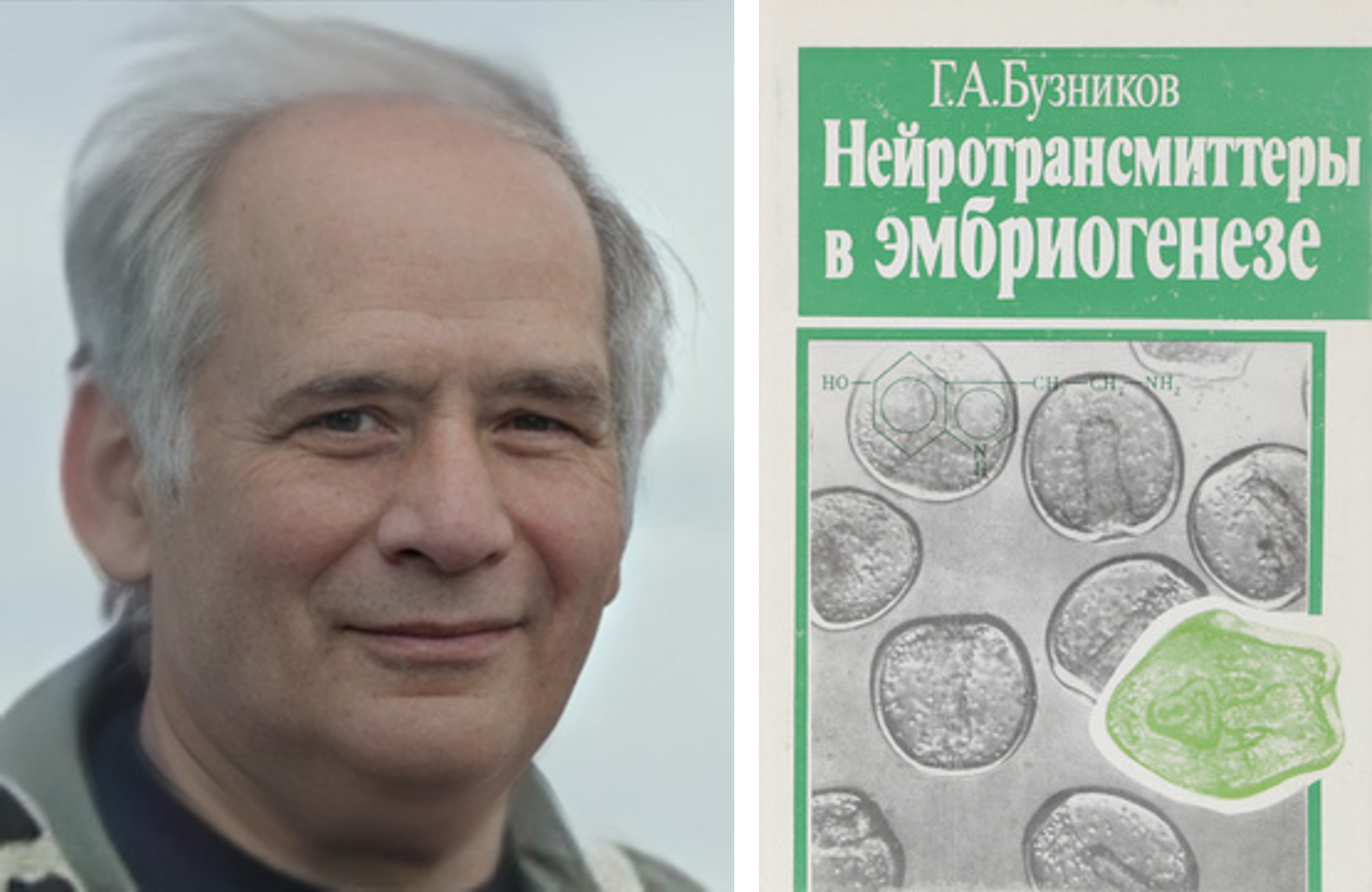
Gennady Alexeevich Buznikov (1931-2012), Doctor of Biological Sciences, Professor, founding father of the studies of the role of the transmitters in embryonic development. Frontpage of his last monography in Russian “Neurotransmitters in embryogenesis” (Moscow, 1987).
Laboratory of Embryophysiology was established in 1977 by professor Gennady A. Buznikov (1931-2012) – father of research into the functions of classic neurotransmitters in prenervous embryogenesis. Buznikov was the first to prove the transmitters' presence and functional activity in the cells of early embryos.
The first evidence of the active role of the transmitters in the prenervous regulations was obtained in the experiments with clutches of White Sea nudibranch mollusks. It was shown that serotonin influences the activity of ciliary motility of embryos having no neurons yet (Koshtoyants et al., 1961). “Consequences were impressive. Buznikov left his previous studies and became fully occupied by “prenervous neurotransmitters.” In 1967 he published the first monography in the field. Later (in 1987), the next was published in Russian and translated in English (1990). Since first Buznikov’s experiments “world science poured into the resulting breakthrough and the glory of the founding father of a new direction in biology is assigned to Buznikov” (Сахаров, 2013).
In subsequent works, Buznikov and his collaborators established that serotonin, catecholamines, and acetylcholine take part in the regulation of essential events of embryogenesis. Among the most notable discoveries made in the Lab of Embryophysiology are as follows:
- the demonstration of the participation of transmitters in the triggering of the cell cycle during cleavage divisions,
- the demonstration of the multiplicity of the transmitters in embryo,
- hypothesis on the intracellular localization of the transmitter receptors in the embryonic cells and later – on the membrane localization and experimental prove of them,
- the experimental evidence of the direct participation of transmitters in blastomere interactions and formation of protosynapse concept,
- demonstration of the interaction of embryonic transmitters with intracellular signal transduction pathways,
- transmitter influence on the morphogenetic movements in the embryo
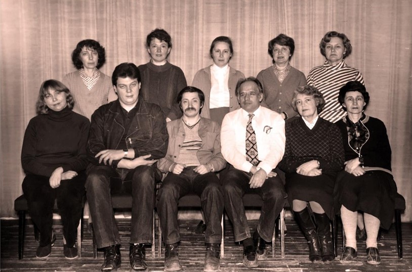
Lab of Embryophysiology, 1984. Upper row from the left: Natalia D. Zvezdina, Olga G. L’vova, Lyudmila A. Mal’chenko, Lidia N. Markova, and Eleonora V. Gusareva. Lower row: Larisa E. Martynova, Nikita G. Grigor’ev, Yuri B. Shmukler, Prof Gennady A. Buznikov, Ninel A. Teplitz, Svetlana A. Ustinova
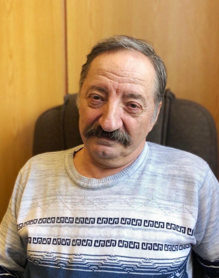
Former Head of the Group, Yuri Shmukler, Doctor of Biological Sciences
A great deal of these experiments was carried out in sea invertebrate embryos, first of all – sea urchins during the expeditions to marine stations of the Barentz Sea and the White Sea, in Vietnam, Yugoslavia, and Italy. As a result, long-lasting and fruitful scientific connections were established.
Since 1996 Prof. Buznikov received the University of North Carolina position but maintained scientific links with the Institute of Developmental Biology and the Lab organized by him. His collaborators continued the studies of transmitters in the early development of mammals, electrophysiological research of membrane transmitter receptors of sea urchins, studied the functional activity of transmitters in the regeneration processes, and the generation of ultradian rhythms in mammalian cells (together with Prof. Vsevolod Brodsky). From 2000 to 2023 the former student and collaborator of Prof. Buznikov Yuri B. Shmukler, Doc Bio. Sci., was the Head of the lab. Today molecular biology studies of the role of prenervous transmitters in the mammalian early embryogenesis and oogenesis have developed most actively in the Group of Embryophysiology led by Denis Nikishin, PhD.
- Buznikov G.A. (1967) Low-molecular weight regulators of embryonic development. Moscow. Nauka Publishers (in Russian)
- Buznikov G.A. (1987) Neurotransmitters in embryogenesis. Moscow. Nauka Publishers (in Russian)
- Koshtoyants Kh.S., Buznikov G.A., Manukhin B.N. The possible role of 5-hydroxytryptamine in the motor activity of embryos of some marine gastropods // Comp. Biochem. Physiol. 1961 V.3. №1. P.20—26.
- Sakharov D.A. And sea angels were with us ... // Priroda. 2013. №2(1170). С. 21–29. (in Russian)