About the Lab
The Laboratory of Regenerative Biology is a structural division in IDB RAS that includes 3 subunits (research groups) additionally to core research topic:
1. Regenerative Biology (core research topic, research lead - Yu. Markitantova)
2. Experimental Neurobiology Research Group (research lead - M. Aleksandrova)
3. Molecular Mechanisms of Ontogeny Research Group (research lead - A. Mikaelyan)
4. Embryophysiology Research Group (research lead - D. Nikishin)
Our research focuses on the molecular and genetic mechanisms of tissue and organ development and regeneration in amphibians and mammals at the cellular, tissue and molecular levels. Key issues are related to the strategies for regenerative responses: cell dedifferentiation and / or activation of cells with stem properties, proliferation of progenitor cells, epithelial-mesenchymal transformation; endogenous and exogenous regulation of the behavior of progenitor cells that constitute the key sources of regeneration.
In amphibians, the retina and lens (the most actively studied models of development and regeneration in our laboratory) are restored as a result of the phenomenon of transdifferentiation (reprogramming in vivo) of pigment epithelial cells and the iris, respectively. We study the molecular participants that control the retinal pigment epitelium (RPE) cell reprogramming in vivo at the level of the local cellular microenvironment (signaling cascades, growth factors, small molecules and transcription factors) and organism factors that can regulate pathological or regenerative processes.
Damage to the human retinal pigment epithelium leads to biochemical and structural disorders (fibrosis, degenerative processes in the retina), including loss of vision (proliferative vitreoretinopathy, etc.). An important aspect of our research is related to the development of methods for reprogramming the RPE to the photoreceptor cells in the mammalian RPE.
The molecular mechanisms of the epithelial-mesenchymal transition that underlies pathology and carcinogenesis in many tissues and organs are also studied by in vivo and in vitro models and experimental approaches.
Keywords: regeneration of tissues and organs, progenitor cells, retinal pigment epithelium, reprogramming in vivo, transdifferentiation, embryogenesis, eye retina morphogenesis, ECM remodeling, transcription factors, signaling pathways, epithelial-mesenchymal transformation, development of methods against fibrosis in the tissues, scaffolds and methods of cell-free therapy
Members of Regenerative Biology Lab
Regenerative Biology

Yuliya Markitantova
Head of the Lab
Chief researcher, PhD
Research interests: strategies for regeneration of the eye tissues, molecular mechanisms of retinal pigment epithelium cell reprogramming, epithelial-mesenchymal transition, regenerative medicine
Eleonora Grigoryan
Chief researcher, PhD, Doctor of Biological Sciences
Research interests: cellular and molecular mechanisms of organ and tissue regeneration in vertebrates

Vladimir Simirskii
Senior researcher, PhD
Research interests: strategies for and molecular mechanisms of eye tissue regeneration

Anna Shabarina
Researcher, PhD
Research interests: cell nucleus structure and chromatin organization
Valentina Poplinskaya
Research assistant, PhD
Specialist in histology and histochemical analysis

Yulia Novikova
Research assistant, PhD
Specialist in organotypic cell culture and histology
Experimental Neurobiology Research Group

Maria Alexandrova
Head of the Group
Chief researcher, PhDDoctor of Biological Sciences
Research interests: neurogenesis in vertebrates, neural stem cells, stem and progenitor cells plasticity regulation, mechanisms of neural tissue regeneration, transplantation
Alla Kuznetsova
Senior researcher, PhD
Research interests: extracellular matrix, decellularized extracellular matrix, regenerative medicine, cell-free therapy; epithelial-mesenchymal transition; cancer-associated fibroblasts; tumor microenvironment, tumor-stromal interactions, molecular mechanisms

Lubov Rjanova
Researcher, PhD
Research interests: transdifferentiation and reprogramming of mammalian and human retinal pigment epithelium cells in vitro, regenerative medicine
Elena Shafei
Research assistant
Research interests: plasticity of retinal pigment epithelial cells and its regulation in vitro, regenerative medicine
Molecular Mechanisms of Ontogeny Research Group
Arsen Mikaelyan
Head of the Group
Senior researcher, PhD
Research interests: cellular and molecular-genetic mechanisms of liver regeneration, fibrosis, carcinogenesis
Natalya Dashenkova
Researcher, PhD
Research interests: molecular mechanisms of liver regeneration, fibrosis, carcinogenesis
Embryophysiology Research Group
Denis Nikishin
Head of the group
Senior Researcher, PhD
Field of interests: molecular mechanisms of transmitter regulations of the early embryogenesis and oogenesis.
Yulia Nikishina
Researcher, PhD
Field of interests: the role of catecholamines in development and ovarian function.
Nina Alyoshina
Junior researcher, PhD
Field of interests: regulation of oogenesis and folliculogenesis by serotonin.
Lyudmila Mal’chenko
Researcher-Engineer
Specialist in cell culture, immunohistochemical, and radioisotope analysis.
Anna Dubrovskaya
PhD Candidate
Thesis research is devoted to studying the role of L-DOPA in polycystic ovarian syndrome.
Research directions
Molecular mechanisms of RPE cell reprogramming, fate of the stem-like neuroblasts and retina morphogenesis during regeneration in lower vertebrates
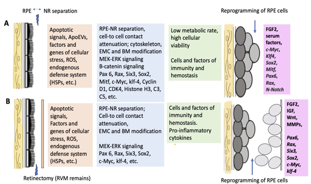
Fig. 1 A range of known cellular and molecular factors involved in the initiation of retinal regeneration and RPE reprogramming in Urodela (A) and Anura (B) (Markitantova, Grigoryan, 2023).
The subject of our research is the retina of the newts Pleurodeles waltl (Urodela) which is capable of complete retinal restoration after its detachment or removal. In this regard, the newt retina is a unique model system for the study of cell fate, the patterns of cell proliferation, apoptosis and differentiation, the mechanisms of morphogenesis in development and regeneration, as well as cell function in health and pathology. It has been shown that the proliferation of a transient neuroblast-like pool of cells, generated from the retinal pigment epithelium (RPE) cells by genome reprogramming, provides retinal regeneration in the adult newt. In retina development, the neuroblast cell population is also a source for generating diverse progenies for all types of neuronal and glial cells. Using morphological, molecular and immunochemical methods, we have identified the pattern of retinal cell type differentiation and molecular regulation of retinal morphogenesis during development and regeneration in the newt P. waltl. We have found genes encoding transcription factors and signaling proteins expressed from the early stages of neuroblasts formed as a result of reprogramming RPE and later – in differentiating neurons of the regenerating retina. Among them are Fgf2, Wntb2, Bmp4, Pax6, Sox2, Rx, Six3, Prox1, Pitx1-3, Otx2 and others. The results indicate that retinal morphogenesis occurs regardless of the origin of progenitor cells (neuroblasts) during development and regeneration under the control of evolutionary conservative regulatory genes. They are involved in the specific gene networks which coordinate cells-type differentiation and eye morphogenesis in vertebrates. In the ciliary growth zone of the newt eye, we have also found the regulatory factors Fgf2, Pax6, Sox2, Rx, Six3, Prox1, Otx2, Pitx1-3 and Gnl3 that are involved in the maintaining of undifferentiated «stem-like» cells. These cells represent the growth and regenerative potential for the retina, along with the RPE cells. Our data also indicate the existence of taxonomic differences.
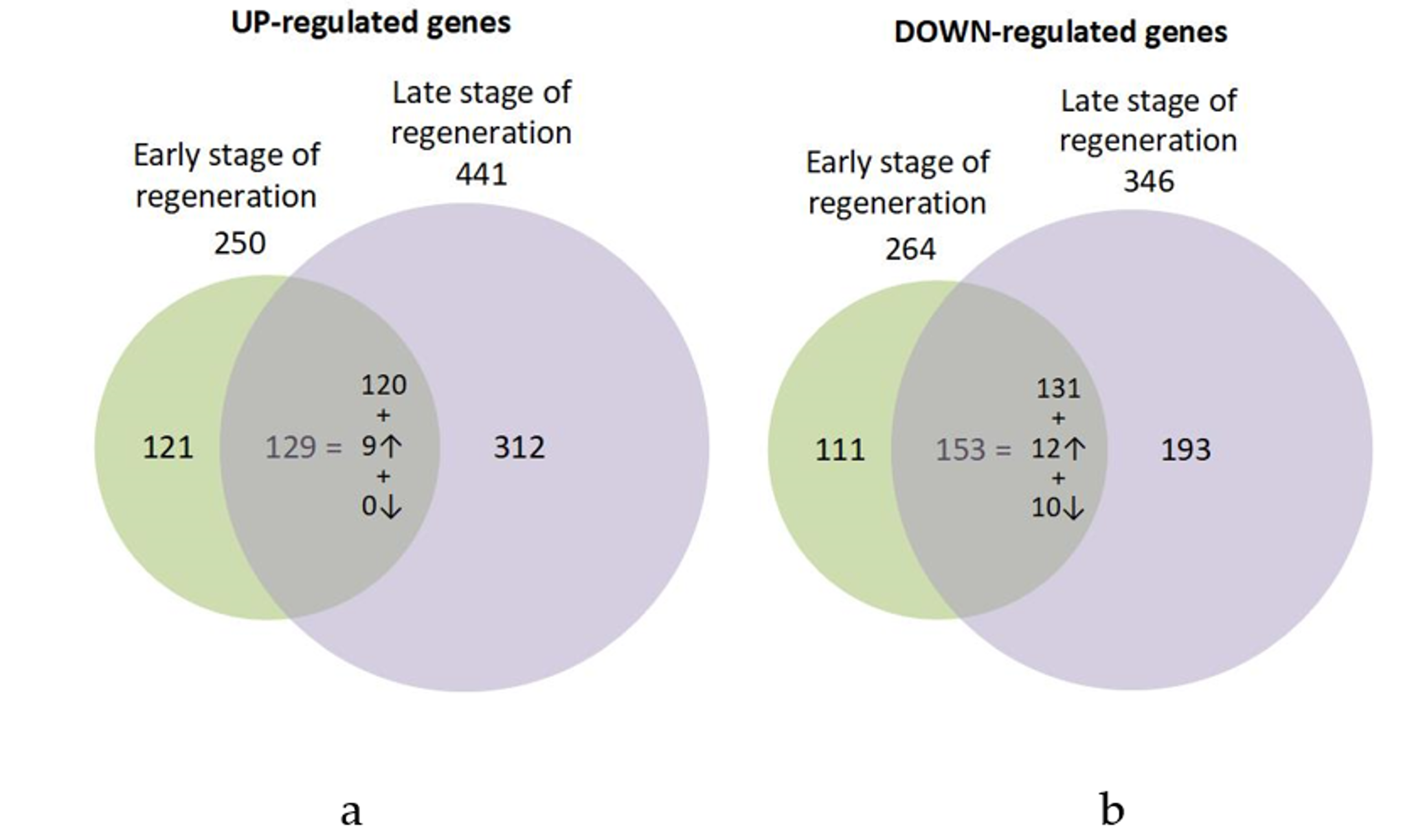
Fig. 2 Transcriptome analysis. Venn diagram of the up- (a) and down-regulated (b) genes in the early and late stages of RPE cell reprogramming during retinal regeneration in the newt P. waltl. The expressions in the regions of intersection of the sets demonstrate the common differentially expressed genes for both stages of regeneration and the proportion of genes with similar levels of differential expression, with a level increased at a later stage twofold or more or decreased twofold or more. The direction of the arrows indicates an increase or decrease in differential expression (Markitantova et al., 2023).
The results obtained on the unique model of the RPE reprogramming in the adult newt during retinal regeneration in terms of the expression of regulatory genes confirm the hypothesis about the “switching of genes work” as a key phenomenon of the process of cell conversion, previously laid down by Professor O.G. Stroeva and Professor V.I. Mitashov (founder of the Laboratory of the Problems of Regeneration). Currently, our research is aimed at searching for key components of endogenous protective systems (transcription factors, etc.) of retinal pigment epithelial cells that allow/block regeneration due to cell conversion, and their phylogenetic characterization. The long-term research directions focusing on retinal regeneration mechanisms in the newts could be briefly summarized as follows: from the study of macromolecular syntheses to the analysis of single gene expression, transcriptomic changes and signaling pathways, and finally work on proteomics and epigenetics.
Selected publications
- Markitantova Y.V., Grigoryan E.N. Cellular and Molecular Triggers of Retinal Regeneration in Amphibians // Life-Basel. 2023. Vol. 13(10). Art. No. 1981. DOI: 10.3390/life13101981.
- Markitantova Y., Fokin A., Boguslavsky D., Simirskii V., Kulikov A. Molecular Signatures Integral to Natural Reprogramming in the Pigment Epithelium Cells after Retinal Detachment in Pleurodeles waltl // Int. J. Mol. Sci. 2023. Vol. 24. Art.no 16940.DOI: 10.3390/ ijms242316940.
- Markitantova Yu.V., Simirskii V.N. Conservatism and Variability of the Antioxidant Defense System in the Retinal Pigment Epithelium of Vertebrates Journal of Evolutionary Biochemistry and Physiology. 2023. No. 3, Vol. 59, pp. 655–675 https://doi.org/10.1134/S0022093023030018
- Markitantova Y., Simirskii V. Endogenous and Exogenous Regulation of Redox Homeostasis in Retinal Pigment Epithelium Cells: An Updated Antioxidant Perspective // Int. J. Mol. Sci. 2023. 24 10776. DOI: 10.3390/ijms241310776. Published: 28 June 2023.
- Grigoryan E.N. Study of natural longlife juvenility and tissue regeneration in caudate amphibians and potential application of resulting data in biomedicine // Journal of Developmental Biology. 2021. Vol. 9. Is. 1. Art. No 2. P. 1-19. DOI: 10.3390/jdb9010002.
- Grigoryan E.N., Markitantova Y.V. Molecular Strategies for Transdifferentiation of Retinal Pigment Epithelial Cells in Amphibians and Mammals In Vivo // Russian journal of Developmental biology. 2021. Vol. 52. Is. 4. P. 220-243. DOI 10.1134/S1062360421040032.
- Grigoryan E.N. Self-Organization of the Retina during Eye Development, Retinal Regeneration In Vivo, and in Retinal 3D Organoids In Vitro // Biomedicines. 2022. Vol. 10. Art. no 1458. DOI: 10.3390/biomedicines10061458
- Markitantova Yu.V., Simirskii V.N. Role of the Redox System in Initiation of a Regenerative Response of Neural Eye Tissues in Vertebrates // Russian Journal of Developmental Biology. 2020. Vol. 51. Is. 1. P. 16-30. DOI: 10.1134/S106236042001004X.
- Markitantova Y.V., Novikova Y.P., Poplinskaya V.A., Grigoryan E.N. Expression of FGF2 and Nucleostemin in Models of Retinal Regeneration in the Newt Under Conditions of 3D Organotypic Culture In vitro//EC Ophthalmology. 2020. V. 11. Is. 1. P. 01-09. DOI:10.31080/ecop.2020.11.00580.
- Grigoryan E.N. Potential Endogenous Cell Sources for Retinal Regeneration in Vertebrates and Humans: Progenitor Traits and Specialization // Biomedicines. 2020. V. 8. P. 208. DOI: 0.3390/biomedicines8070208.
- Markitantova Yu.V., Simirskii V.N. Inherited retinal diseases through the eyes of homeobox genes // Int. J. Mol. Sci. 2020. V. 21. № 5. P. 1602.Doi: 10.3390/ijms21051602
Studies of the mechanisms of transdifferentiation and reprogramming of mammalian and human retinal pigment epithelium cells in vitro
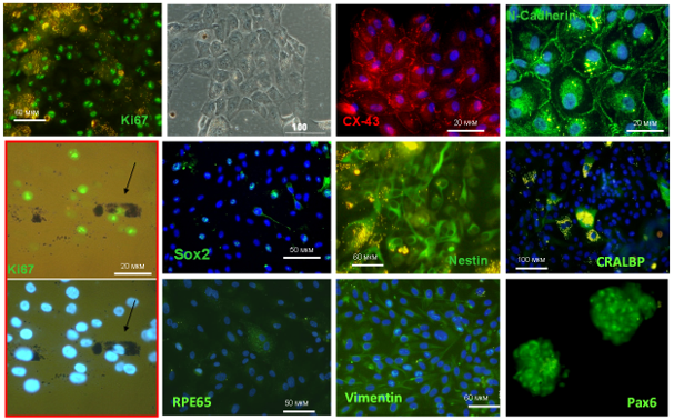
Fig. 3 Expression of marker proteins in dedifferentiated human retinal pigment epithelial cells in vitro.
Regeneration of the retina of vertebrate eyes is due to internal and external reserves. Neuronal degeneration in the human retina is often a consequence of pathological reprogramming of plastic pigment epithelial (RPE) cells capable of differentiation in the mesenchymal and neural directions. Degenerative diseases or injuries to nervous system structures often have no effective treatment. The work of the experimental neurobiology research group (Dr. M.A. Aleksandrova) is aimed at studying the participation of stem and highly plastic cells of the nervous tissue of animals and humans in regeneration processes. Our studies focus on cellular approaches: this includes the regulation of cell reprogramming in vitro, as well as studies on the effect of transplantation of various cell types on the restoration of central nervous system functions. Transdifferentiation and reprogramming of the differentiated retinal pigment epithelium (RPE) are studied using an in vitro model, the human cell line ARPE-19. RPE cells of the adult human eye, when cultivated in vitro, undergo phenotypic conversion towards neural and epithelial differentiation and exhibit stem cell properties. Several signaling proteins involved in changing the nature of differentiation of adult RPE cells and ARPE-19 culture cells have been identified. A decrease in the level of differentiation of adult RPE cells in the early stages of the action of oFGF was established, including: inactivation of the Wnt/β-catenin and Notch signaling, activation of the non-canonical Wnt/PCP signaling and modulation of BMP-signaling. The mechanisms of cell reprogramming and ways to search for molecules that regulate RPE plasticity are currently being studied.
Selected publications
- Shafei E.V., Rzhanova L.A., Novikova Y.P., Kurinov A.M., Grigoryan E.N., Aleksandrova M.A., Kuznetsova А.V. Response of human retinal pigment epithelial cells to the effect of the conditioned media of newt retinal regenerates // Cell and Tissue Biology. 2021. Vol. 15. № 2. P. 135-149. DOI: 10.1134/S1990519X21020085
- Rzhanova L.A., Kuznetsova A.V., Aleksandrova M.A. Reprogramming of differentiated mammalian and human retinal pigment epithelium: current achievements and prospects // Russian Journal of Developmental Biology. 2020. V. 51. №. 4. P. 212–230. DOI: 10.1134/S1062360420040062
- Kuznetsova А.V., Rzhanova L.A., Kurinov A.M., Aleksandrova М.A. Effect of basic fibroblast growth factor on signaling pathways in adult human retinal pigment epithelial cells // Cell and Tissue Biology. 2019. Vol. 13, № 4. P. 292-304. DOI: 10.1134/S1990519X19040059.
- Shafei E.V., Kurinov A.M., Kuznetsova A.V., Aleksandrova M.A. Reprogramming of human retinal pigment epithelial cells under the effect of bFGF in vitro // Bulletin of Experimental Biology and Medicine. 2017. V. 163. 4. P. 574–582). https://doi.org/10.1007/s10517-017-3852-5
- Kuznetsova A.V., Aleksandrova M.A., Kurinov A.M., Chentsova E.V., Makarov P.V. Plasticity of adult human retinal pigment epithelial cells // International Journal of Clinical and Experimental Medicine. 2016. V. 9. № 11. Р. 20892-20906.
Mechanisms of tissue regeneration, fibrosis and cancers
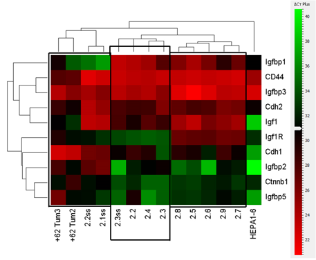
Fig. 4 Cluster analysis of data obtained by real-time PCR on the expression of the studied genes in chemically induced liver hepatocarcinoma.
An elevated plasma level of hyaluronic acid (HA) is a disease marker in liver pathology and other inflammatory disorders. Inhibition of HA synthesis with coumarin 4-methylumbelliferone (4MU) has a beneficial effect in animal models of fibrosis, inflammation, cancer and metabolic syndrome. 4MU is an active compound of approved choleretic drug hymecromone with low bioavailability and a broad spectrum of action. New, more specific and efficient inhibitors of hyaluronan synthases (HAS) are required. We have tested several newly synthesized coumarin compounds and commercial chitin synthesis inhibitors to inhibit HA production in cell culture assay. Coumarin derivative compound VII (10'-methyl-6'-phenyl-3'H-spiro[piperidine-4,2'-pyrano[3,2-g]chromene]-4',8'-dione) demonstrated inhibition of HA secretion by NIH3T3 cells with the half-maximal inhibitory concentration (IC50) = 1.69 ± 0.75 μΜ superior to 4MU (IC50 = 8.68 ± 1.6 μΜ). Inhibitors of chitin synthesis, etoxazole, buprofezin and triflumuron, reduced HA deposition with IC50 of 4.21 ± 3.82 μΜ, 1.24 ± 0.87 μΜ and 1.48 ± 1.44 μΜ, respectively. Etoxazole reduced HA production and prevented collagen fiber formation in the CCl4 liver fibrosis model in mice similar to 4MU. Bioinformatics analysis revealed homology between chitin synthases and HAS enzymes, particularly in the pore-forming domain, containing the proposed site for etoxazole binding.
Our analysis of public databases shows that exposure to 4MU leads to the activation of pathways associated with the cellular stress response through the transcription factors HIF1A, NFkB, NFE2L2, and ATF6. Our study revealed novel biological targets of 4MU, including enzymes, nuclear receptors and transcription factors. A detailed study of the 4MU targets identified will be critical for the synthesis of new drugs for the effective treatment of pathological processes associated with metabolic disorders, inflammation, regeneration and oncogenesis in liver. The transcriptomic analysis performed in our study is consistent with other researchers' findings that nuclear receptors may be the main targets of 4MU. (Research lead - Ph.D. Mikaelyan A.S.)
Selected publications
- Tsitrina A.A, Halimani N., Andreichenko I.N., Sabirov M, Nesterchuk M., Dashenkova N.O., Romanov R., Bulgakova E.V., Mikaelyan A., Kotelevtsev Y. 4-Methylumbelliferone Targets Revealed by Public Data Analysis and Liver Transcriptome Sequencing // International Journal of Molecular Sciences. 2023. Vol. 24. Art. no 2129. DOI: 10.3390/ijms24032129.
- Shashkovskaya V.S., Vetosheva P.I., Shokhina A.G., Aparin I.O., Prikazchikova T.A., Mikaelyan A.S.1, Kotelevtsev Y.V., Belousov V.V., Zatsepin T.S., Abakumova T.O. Delivery of Lipid Nanoparticles with ROS Probes for Improved Visualization of Hepatocellular Carcinoma // Biomedicines. 2023. Vol. 11(7). Art. no. 1783. DOI: 10.3390/biomedicines11071783.
- Abakumova T., Vaneev A., Naumenko V., Shokhina A, Belousov V., Mikaelyan A., Balysheva K., Gorelkin P., Erofeev A., Zatsepin T. Intravital electrochemical nanosensor as a tool for the measurement of reactive oxygen/nitrogen species in liver diseases // J Nanobiotechnology. 2022;20(1):497. DOI: 10.1186/s12951-022-01688-z
- Sapach A.Yu., Sindeeva O.A., Nesterchuk M.V., Tsitrina A.A., Mayorova O.A., Prikhozhdenko E.S., Verkhovskii R.A., Mikaelyan A.S., Kotelevtsev Y.V., Sukhorukov G.B. Macrophage In Vitro and In Vivo Tracking via Anchored Microcapsules // ACS Applied Materials & Interfaces. 2022. https://doi.org/10.1021/ACSAMI.2C12004
- Andreichenko I.N., Tsitrina A.A., Fokin A.V., Gabdulkhakova A.I., Maltsev D.I., Perelman G.S., Bulgakova E.V., Kulikov A.M., Mikaelyan A.S., Kotelevtsev Y.V. 4-methylumbelliferone prevents liver fibrosis by affecting hyaluronan deposition, FSTL1 expression and cell localization // Int. J. Mol. Sci. 2019. V. 20. Is. 24. Р. 6301-6318. DOI:10.3390/ijms20246301.
Development of cell-free scaffolds for regenerative medicine
The influence of extracellular matrix components on the differentiation of periodontal ligament stem cells is studied. The cell-free products that are being developed for use in regenerative medicine can improve the migration, proliferation, differentiation and metabolism of various resident stem cells, which will ensure the regulation of spatially correct their location and will stimulate endogenous regeneration of damaged tissues. Cell-free therapeutic agents leading to the recruitment/homing of endogenous SCs have an advantage in overcoming the limitations and risks associated with the use of cell therapy, including tumor formation, undesirable immune responses and pathogen transmission, and also have significant benefits in production, storage and standardization.
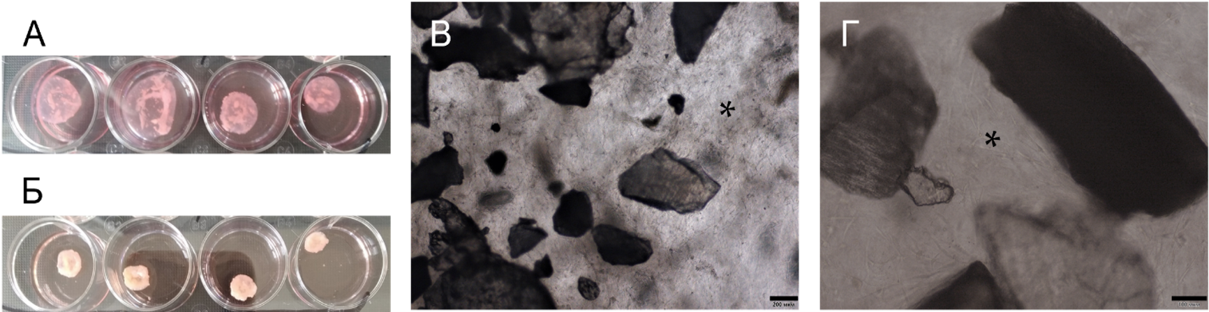
Fig. 5 Bioengineered construct (acellular scaffolds) together with periodontal cells in collagen I hydrogel (3D culture).
Selected publications
- Ivanov A.A., Danilova T.I., Kuznetsova A.V, Popova O.P., Yanushevich O.O. Decellularized Matrix Induced Spontaneous Odontogenic and Osteogenic Differentiation in Periodontal Cells // Biomolecules. 2023. Vol. 13(1). Art. no. 122. DOI: 10.3390/biom13010122.
- Ivanov A.A., Kuznetsova A.V., Popova O.P., Danilova T.I., Latyshev A.V., Yanushevich O.O. Influence of Extracellular Matrix Components on the Differentiation of Periodontal Ligament Stem Cells in Collagen I Hydrogel // Cells. 2023. Vol.12 (19). Art. no 2335. DOI: 10.3390/cells12192335.
Financial support and collaborations
Grants of the Russian Science Foundation
- RSF grant for young scientists. Project No. 19-74-00117 (2019-2021) «Development of technology for growing three-dimensional human and mouse cerebral organoids in chimeric and xenochimeric models in vivo» (Research lead: K.K. Sukhinich)
- Grant of the Russian Science Foundation Project No. 222500835 (2022 – 2023) “Features of the early regenerative response of RPE cells after experimental retinal detachment: study of the relationship between oxidative stress and the purinergic system” (Research lead: Yu.V. Markitantova)
Grants of the Russian Foundation for Basic Research
- Grant RFBR Project № 16-04-01114 (2016-2018) Study of the role of components of the purinergic signaling system in the regulation of morphogenesis of eye tissue in lower and higher vertebrates in ontogenesis, regeneration and conditions of experimentally induced pathology (Research lead: Yu.V. Markitantova)
- Russian Foundation for Fundamental Research, Project № 14-04-00184 (2014-2016), (Research lead: E.N. Grigoryan)
Agreement on scientific cooperation
- Skoltech-IBR Research Agreement No. 10.04.18 “Search for hyaluronan synthase inhibitors with hepatoprotective and antifibrotic activity and study of the mechanism of their action” (Research lead: A.S. Mikaelyan)
Collaborations
- Department of Embryology of Biological Faculty, M.V. Lomonosov Moscow State University
- State budgetary healthcare institution of the Moscow region Moscow Regional Research Clinical Institute named after M. F. Vladimirsky
- Federal State Budgetary Institution of Higher Education "Moscow State Medical and Dental University named after A.I. Evdokimov, Laboratory of Molecular Cell Pathology
- Skolkovo Institute of Science and Technology (Skoltech)
General publications
- Markitantova Y.V., Grigoryan E.N. Cellular and Molecular Triggers of Retinal Regeneration in Amphibians // Life-Basel. 2023. Vol. 13(10). Art. No. 1981. DOI: 10.3390/life13101981
- Markitantova Y., Fokin A., Boguslavsky D., Simirskii V., Kulikov A. Molecular Signatures Integral to Natural Reprogramming in the Pigment Epithelium Cells after Retinal Detachment in Pleurodeles waltl // Int. J. Mol. Sci. 2023. Vol. 24. Art.no 16940.DOI: 10.3390/ ijms242316940. 29 November 2023
- Markitantova Y.V., Grigoryan E.N. Cellular and Molecular Triggers of Retinal Regeneration in Amphibians // Life-Basel. 2023. Vol. 13(10). Art. No. 1981. DOI: 10.3390/life13101981.
- Markitantova Y., Simirskii V. Endogenous and Exogenous Regulation of Redox Homeostasis in Retinal Pigment Epithelium Cells: An Updated Antioxidant Perspective // Int. J. Mol.Sci. 2023. 24 10776. DOI: 10.3390/ijms241310776. Published: 28 June 2023.
- Markitantova Yu.V., Simirskii V.N. Conservatism and Variability of the Antioxidant Defense System in the Retinal Pigment Epithelium of Vertebrates Journal of Evolutionary Biochemistry and Physiology. 2023. No. 3, Vol. 59, pp. 655–675 https://doi.org/10.1134/S0022093023030018
- Musayev-Galbinur P., Markitantova Yu., Babayev Kh., Akberova S. Hypoxia-induced apoptosis of eyeball cells//Journal of Life Sciences & Biomedicine. 2023. Vol. 5(78). No 1. P. 38-41. DOI: 10.5281/zenodo.8004205.
- Grigoryan E.N. Impact of Microgravity and Other Spaceflight Factors on Retina of Vertebrates and Humans In Vivo and In Vitro // Life 2023. 13. 1263.
- Ivanov A.A., Danilova T.I., Kuznetsova A.V, Popova O.P., Yanushevich O.O. Decellularized Matrix Induced Spontaneous Odontogenic and Osteogenic Differentiation in Periodontal Cells // Biomolecules. 2023. Vol. 13(1). Art. no. 122. DOI: 10.3390/biom13010122.
- Ivanov A.A., Kuznetsova A.V., Popova O.P., Danilova T.I., Latyshev A.V., Yanushevich O.O. Influence of Extracellular Matrix Components on the Differentiation of Periodontal Ligament Stem Cells in Collagen I Hydrogel // Cells. 2023. Vol.12 (19). Art. no 2335. DOI: 10.3390/cells12192335.
- Shashkovskaya V.S., Vetosheva P.I., Shokhina A.G., Aparin I.O., Prikazchikova T.A., Mikaelyan A.S, Kotelevtsev Y.V., Belousov V.V., Zatsepin T.S., Abakumova T.O. Delivery of Lipid Nanoparticles with ROS Probes for Improved Visualization of Hepatocellular Carcinoma // Biomedicines. 2023. Vol. 11(7). Art. no. 1783. DOI: 10.3390/biomedicines11071783.
- Tsitrina A.A, Halimani N., Andreichenko I.N., Sabirov M, Nesterchuk M., Dashenkova N.O., Romanov R., Bulgakova E.V., Mikaelyan A., Kotelevtsev Y. 4-Methylumbelliferone Targets Revealed by Public Data Analysis and Liver Transcriptome Sequencing // International Journal of Molecular Sciences. 2023. Vol. 24. Art. no 2129. DOI: 10.3390/ijms24032129.
- Osidak E.O., Andreev A.Y., Avetisov S.E., Voronin G.V., Surnina Z.V., Zhuravleva A.V., Grigoriev T.E., Krasheninnikov S.V., Sukhinich K.K., Zayratyants O.V., Domogatsky S.P. Corneal Stroma Regeneration with Collagen-Based Hydrogel as an Artificial Stroma Equivalent: A Comprehensive In Vivo Study // Polymers. 2022. Vol. 14. Is. 19. Art. no 4017. doi.org/10.3390/polym14194017
- Abakumova T., Vaneev A., Naumenko V., Shokhina A, Belousov V., Mikaelyan A., Balysheva K., Gorelkin P., Erofeev A., Zatsepin T. Intravital electrochemical nanosensor as a tool for the measurement of reactive oxygen/nitrogen species in liver diseases // J Nanobiotechnology. 2022;20(1):497. DOI: 10.1186/s12951-022-01688-z
- Sapach A.Yu., Sindeeva O.A., Nesterchuk M.V., Tsitrina A.A., Mayorova O.A., Prikhozhdenko E.S., Verkhovskii R.A., Mikaelyan A.S., Kotelevtsev Y.V., Sukhorukov G.B. Macrophage In Vitro and In Vivo Tracking via Anchored Microcapsules // ACS Applied Materials & Interfaces. 2022. https://doi.org/10.1021/ACSAMI.2C12004
- Grigoryan E.N. Self-Organization of the Retina during Eye Development, Retinal Regeneration In Vivo, and in Retinal 3D Organoids In Vitro // Biomedicines. 2022. Vol. 10. Art. no 1458. DOI: 10.3390/biomedicines10061458
- Shafei E.V., Rzhanova L.A., Novikova Y.P., Kurinov A.M., Grigoryan E.N., Aleksandrova M.A., Kuznetsova А.V. Response of human retinal pigment epithelial cells to the effect of the conditioned media of newt retinal regenerates // Cell and Tissue Biology. 2021. Vol. 15. № 2. P. 135-149. DOI: 10.1134/S1990519X21020085
- Panova I.G., Sukhova Y.V., Tatikolov A.S., Levin P.P., Ivanets T.Y. Antioxidants in the Vitreous Body of the Eye of Human Fetuses // Biology Bulletin. 2021. Vol. 48. Is. 6. P. 662-666. DOI: 10.1134/S1062359021050113.
- Grigoryan E.N., Markitantova Y.V. Molecular Strategies for Transdifferentiation of Retinal Pigment Epithelial Cells in Amphibians and Mammals In Vivo // Russian journal of Developmental biology. 2021. Vol. 52. Is. 4. P. 220-243. DOI 10.1134/S1062360421040032.
- Grigoryan E.N. Study of natural longlife juvenility and tissue regeneration in caudate amphibians and potential application of resulting data in biomedicine // Journal of Developmental Biology. 2021. Vol. 9. Is. 1. Art. No 2. P. 1-19. DOI: 10.3390/jdb9010002.
- Sukhinich K.K., Aleksandrova M.A. Cerebral Organoids: A Model of Brain Development // Russian Journal of Developmental Biology. 2020. V. 51. Is. 4. P. 231-245. DOI: 10.1134/S1062360420040074.
- Grigoryan E.N. Potential Endogenous Cell Sources for Retinal Regeneration in Vertebrates and Humans: Progenitor Traits and Specialization // Biomedicines. 2020. V. 8. P. 208. DOI: 0.3390/biomedicines8070208.
- Markitantova Yu.V., Simirskii V.N. Role of the Redox System in Initiation of a Regenerative Response of Neural Eye Tissues in Vertebrates // Russian Journal of Developmental Biology. 2020. Vol. 51. Is. 1. P. 16-30. DOI: 10.1134/S106236042001004X.
- Markitantova Yu.V., Simirskii V.N. Inherited retinal diseases through the eyes of homeobox genes // Int. J. Mol. Sci. 2020. V. 21. № 5. P. 1602.Doi: 10.3390/ijms21051602
- Novikova Yu., Grigoryan E.N. Early Appearance of Aging Signs in the Retinal Pigment Epithelium in Young Albino Rats // Russian Journal of Developmental Biology. 2020. V. 51. No. 6. Р. 377–386. DOI: 10.1134/S1062360420060065.
- Markitantova Y.V., Novikova Y.P., Poplinskaya V.A., Grigoryan E.N. Expression of FGF2 and Nucleostemin in Models of Retinal Regeneration in the Newt Under Conditions of 3D Organotypic Culture In vitro//EC Ophthalmology. 2020. V. 11. Is. 1. P. 01-09. DOI:10.31080/ecop.2020.11.00580.
- Rzhanova L.A., Kuznetsova A.V., Aleksandrova M.A. Reprogramming of differentiated mammalian and human retinal pigment epithelium: current achievements and prospects // Russian Journal of Developmental Biology. 2020. V. 51. №. 4. P. 212–230. DOI: 10.1134/S1062360420040062
- Novikova Yu.P., Poplinskaya V.A., Grigoryan E.N. Organotypic Culturing as a Way to Study Recovery Opportunities of the Eye Retina in Vertebrates and Humans // Russian Journal of Developmental Biology. 2020. V. 51. Is. 1. P. 31-44. DOI: 10.1134/S1062360420010063.
- Sukhinich K.K., Dashinimaev E.B., Vorotelyak E.A., Aleksandrova M.A. Regenerative Effects and Development Patterns of Solid Neural Tissue Grafts Located in Gelatin Hydrogel Conduit for Treatment of Peripheral Nerve Injury // Plastic and Reconstructive Surgery. Global Open. 2020. Art: GOX-D-19-00596. DOI: 10.1097/GOX.0000000000002610.
- Panova I.G., Sukhova Y.V., Tatikolov A.S., Ivanets T.Yu. Bilirubin in the Vitreous Body of the Eye of Human Fetuses // Bull Exp Biol Med. 2020. V. 170. No. 1. P. 98-100. DOI: 10.1007/s10517-020-05012-3.
- Andreichenko I.N., Tsitrina A.A., Fokin A.V., Gabdulkhakova A.I., Maltsev D.I., Perelman G.S., Bulgakova E.V., Kulikov A.M., Mikaelyan A.S., Kotelevtsev Y.V. 4-methylumbelliferone prevents liver fibrosis by affecting hyaluronan deposition, FSTL1 expression and cell localization // Int. J. Mol. Sci. 2019. V. 20. Is. 24. Р. 6301-6318. DOI:10.3390/ijms20246301.
- Kuznetsova А.V., Rzhanova L.A., Kurinov A.M., Aleksandrova М.A. Effect of basic fibroblast growth factor on signaling pathways in adult human retinal pigment epithelial cells // Cell and Tissue Biology. 2019. Vol. 13, № 4. P. 292-304. DOI: 10.1134/S1990519X19040059.
- Grigoryan E.N., Radugina E.A. Behavior of Stem-Like Cells, Precursors for Tissue Regeneration in Urodela, Under Conditions of Microgravity // Stem cells and development. 2019. V. 28. Is. 7. P. 423-437. DOI: 10.1089/scd.2018.0220
- Namestnikova D.D., Gubskiy I.L., Salikhova D.I., Leonov G.E., Sukhinich K.K., Melnikov, P.A., Vishnevskiy, D.A., Cherkashova, E.A., Gabashvili, A.N., Bukharova, T.B., Burunova, V.V., Fatkhudinov, T.Kh., Chekhonin, V.P., Gubsky, L.V., Kiselev, S.L., Goldstein, D.V., Yarygin, K.N. Therapeutic efficacy of intra-arterial administration of induced pluripotent stem cells-derived neural progenitor cells in acute experimental ischemic stroke in rats // Vestnik Transplantologii i Iskusstvennykh Organov. 2019. V. 21(1). P. 153-164. DOI: 10.15825/1995-1191-2019-1-153-164
- Radugina E.A., Poplinskaya V.A., Markitantova Y.V., Grigoryan E.N., Almeida E.A.C., Blaber E. Exposure to microgravity for 30 days onboard bion m1 caused muscle atrophy and impaired regeneration in murine femoral quadriceps // Life Sciences in Space Research. 2018. V. 16. P. 18-25. DOI: 10.1016/j.lssr.2017.08.005.
- Markitantova Y.V., Akberova S.I., Ryabtseva A.A., Stroeva O.G. The Effect of para-aminobenzoic acid on apoptosis processes in the adult rat conjunctiva and corneal epithelium in vivo after hypobaric hypoxia // Biology Bulletin. 2018. V. 45. N 3. P. 226-234. DOI: 10.1134/S1062359018020061.
- Sukhinich KK, Aleksandrova MA. Individual Peculiarities of the Development and Differentiation of Embryonic Neocortex Transplants in Intact Adult Mouse Brain // Bull Exp Biol Med. 2018. DOI: 10.1007/s10517-018-4303-7.
- Panova I.G., Bezzubenko Yu.V., Tatikolov A.S., Poltavtseva R.A., Ivanets T.Yu., Sukhikh G.T. Alpha-Fetoprotein in Retina and Lens of the Human Eye at Early Stages of Prenatal Development // J Evol Biochem Physiol. 2018. V. 54. No. 2. P. 119—122.
- Panova I.G., Yakovleva M.A., Tatikolov A.S., Kononikhin A.S., Feldman T.B., Poltavtseva R.A., Nikolaev E.N., Sukhikh G.T., Ostrovsky M.A. Lutein and its oxidized forms in eye structures throughout prenatal human development // Experimental Eye Research. 2017. V. 160. P. 31-37.
- Shafei E.V., Kurinov A.M., Kuznetsova A.V., Aleksandrova M.A. Reprogramming of human retinal pigment epithelial cells under the effect of bFGF in vitro // Bulletin of Experimental Biology and Medicine. 2017. V. 163. 4. P. 574–582). https://doi.org/10.1007/s10517-017-3852-5
- Grigoryan E.N., Markitantova Y.V. Cellular and Molecular Preconditions for Retinal Pigment Epithelium (RPE) Natural Reprogramming during Retinal Regeneration in Urodela // Biomedicines. 2016. 4 (4). 28: 1-18.
- Kuznetsova A.V., Aleksandrova M.A., Kurinov A.M., Chentsova E.V., Makarov P.V. Plasticity of adult human retinal pigment epithelial cells // International Journal of Clinical and Experimental Medicine. 2016. V. 9. № 11. Р. 20892-20906.
- Akberova S.I., Markitantova Yu.V., Ryabtseva A.A., Stroeva O.G. Hypoxia as a pathogenic factor affecting eye tissue: selective apoptotic damage to the conjunctiva and anterior corneal epithelium // Reports of the Academy of Sciences (biochemistry, biophysics and molecular biology). Doklady AN (biohimija, biofizika i molekuljarnaja biologija. 2016. 467(6). 718–720. DOI:
10.7868/S0869565216120240 - Panova I.G. Markitantova Y.V., Smirnova Y.A., Zinovieva R.D.
Molecular-genetic mechanisms of cornea morphogenesis // Biology Bulletin. 2015. V. 42. № 2. P. 83-91. - Markitantova Yu.V., Zinovieva R.D. Intracellular localization of transcription factor PROX1 in the human retina in ontogeny // Biology Bulletin 2014. vol. 41(issue 2). P. 103 – 108 DOI:10.1134/S106235901402006X.
- Markitantova Yu.V., Zinovieva R.D. Expression of nucleostemin in proliferating and differentiating cells of the human retina during prenatal development // Dokl Biol Sci. 2012. 445:244-6. doi: 10.1134/S0012496612040084.
- Grigoryan E.N., Novikova Y.P., Gancharova O.S., Kilina O.V., Philippov P.P. New antioxidant SkQ1 is an effective protector of rat eye retinal pigment epithelium and choroid under conditions of long-term organotypic cultivation // Advances in Aging Research. 2012. V. 1. N. 2. P. 31-37.
- Markitantova, Y.V.; Avdonin, P.P.; Grigoryan, E.N.; Zinovieva, R.D. Identification of the pitx1 embryogenesis regulatory gene in a regenerating newt retina // Dokl. Biol. Sci. 2010. 435. 421–424.
- Avdonin, P.P.; Grigoryan, E.N.; Markitantova, Y.V. Transcriptional factor Pitx2: Localization during triton retina regeneration. Biol. Bull. 2010. 37, 231–235.
- Avdonin, P.P.; Grigoryan, E.N.; Markitantova, Y.V. Transcriptional factor Pitx2: Localization during triton retina regeneration // Biol. Bull. 2010. 37. 231–235.
- Desai VD, Wang Y, Simirskii VN, Duncan MK. CD44 expression is developmentally regulated in the mouse lens and increases in the lens epithelium after injury. Differentiation. 2010. V.79. №2. P.111-119.
- Avdonin, P.P.; Markitantova, Y.V.; Zinov’eva, R.D.; Mitashov, V.I. Expression of regulatory genes Pax6, Otx2, Six3, and FGF2 during newt retina regeneration // Biol. Bull. 2008. 35. 355–361.
- Chen X., Patel T.P., Simirskii V.N., Duncan M.K. PCNA interacts with Prox1 and represses its transcriptional activity. Mol. Vis. Vol. 2008. 14. P. 2076-2086.
- Chiba, C.; Mitashov, V.I. Cellular and molecular events in the adult newt retinal regeneration. In Strategies for Retinal Tissue Repair and Regeneration in Vertebrates: From Fish to Human; Chiba, C., Ed.; Research Signpost: Kerala, India, 2007; pp. 15–33.
- Panova I.G., Sharova N.P., Dmitrieva S.B., Poltavtseva R.A., Sukhikh G.N., Tatikolov A.S. Use of a cyanine dye as a probe for albumin and collagen in the extracellular matrix // Analyt. Biochem. 2007. V. 361. P. 183-189.
- Simirskii V.N., Wang Y., Duncan M.K. Conditional deletion of beta1-integrin from the developing lens leads to loss of the lens epithelial phenotype // Dev. Biol. Vol. 2007.306. №2. P. 658-668.
- Markitantova, Y.V.; Makar’ev, E.O.; Smirnova, Y.A.; Zinov’eva, R.D.; Mitashov, V.I. Analysis of the expression pattern of regulatory genes Pax6, Prox1, and Six3 during regeneration of eye structures in the newt // Biol. Bull. 2004. 31. 428–436.
- Stroeva O.G., Panova I.G. Regulation of mitotic activity in the cornea of rats with the protective and therapeutic effects of para‑aminobenzoic acid in experiments with X‑ray irradiation // Proceedings of the RAS. ser. Biological
Izvestija RAN. ser. Biologicheskaja. 1999; 5:613–616 - Grigoryan E.N., Anton H.J., Mitashov V.I. Microgravity effects on neural retina regeneration in the newt // Adv. Space Res. 1998. V. 22. N. 2. P. 293-301.
- Mitashov V.I. Mechanisms of retina regeneration in urodeles // Int. J. Dev. Biol. 1996. 40. 833–844.
- Grigorian, E.N.; Ivanova, I.P.; Poplinskaia, V.A. The discovery of new internal sources of neural retinal regeneration after its detachment in newts: Morphological and quantitative research // Izv. Akad. Nauk. Ser. Biol. 1996. 3. 319–332.
- Mitashov V.I., Arsanto J.-P., Markitantova Y.V., Thouveny Y. Remodeling processes during neural retinal regeneration in adult urodeles: an immunohistochemical survey. International Journal of Developmental Biology // 1995. V. 39 (6). P. 993-1003.
- Mitashov V.I.; Grigorian E.N.; Chernova N.V. Radioautographic study of nonhistone protein synthesis in the process of lens and retina regeneration in adult tritons // Ontogenez. 1983. 14. 390–397.
- Grigoryan E.N.; Mitashov V.I. Cultivation of retinal pigment epithelium in the cavity of lensectomized newt eye // Ontogenez 1985. 16. 34–43.
- Mitashov V.I.; Starostin V.I.; Sludskaia A.I.; Parshina E.F. 3H-thymidine incorporation into the macrophages in the process of eye regeneration in adult tritons // Ontogenez. 1979. 10. 365–371.
- Grigoryan E.N.; Mitashov V.I. An autoradiographic assay of proliferation and melanin synthesis in the pigment epithelium cells during the eye regeneration in newts // Ontogenez. 1979. 10. 137–144.
History of the Lab
The Laboratory of Regeneration Problems was established in 1985 by Professor Victor Ivanovich Mitashov, Doctor of Biological Sciences, who headed the laboratory until 2007. Prof. Mitashov discovered retinal regeneration as a result of RPE cell transdifferentiation (reprogramming in vivo) in the adult newt P. waltl and initiated the studies on the molecular mechanisms of this phenomenon, a key research direction in the field of developmental biology. From 2007 to 2019, Eleonora Norairovna Grigoryan, Doctor of Biological Sciences, headed the laboratory and supervised the research directions.

From the left to right: Professor Victor Ivanovich Mitashov, Doctor of Biological Sciences (3.06.1937-24.04.2007), a founding father of the studies of the cellular and molecular mechanisms of retinal pigment epithelium cell transdifferentiation in adult newt Pleurodeles waltl; Ina Georgievna Panova, Doctor of Biological Sciences; and Olga Georgievna Stroeva, Doctor of Biological Sciences (1925-2021), who founded the research direction in the field of comparative embryological studies of vertebrate eye development (Lopashov G.V., Stroeva O.G. Morphogenesis of the Vertebrate eye // Advances in Morphogenesis. Academic Press: N.Y. USA; London. UK, 1961. V. 1. P. 231–377).
In the retina of adult newts, along with the known cellular sources of its regeneration (pigment epithelium and progenitor cells of the growth zone), the proliferating cells (bipolares) were discovered using 3H-thymidine and BrdU (Dr. E.N. Grigoryan).
Long-term studies of the cellular, molecular and epigenetic mechanisms of tissue morphogenesis in normal regeneration and under modulation of the gravitational load made a contribution to the development of gravitational biology and biomedical treatment.
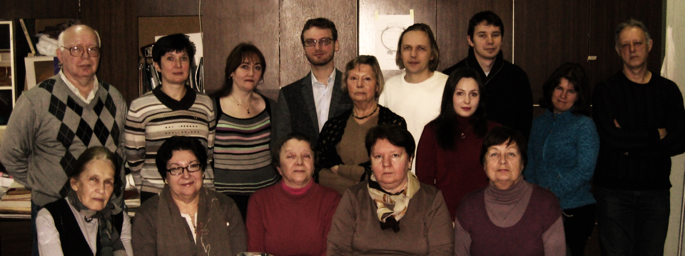
Laboratory of Regeneration Problems, 2016.
Upper row, from the left: Simirskii V.N., Kuznetsova A.V., Markitantova Yu.V., Suchinich K.K, Aleksandrova M.A., Kurinov A.M., Dashenkova N.O., Avdonin P.P., Novikova Yu.A., Mikaelyan A.S. Lower row: Stroeva O.G., Grigoryan E.N., Poplinskay V.A., Panova I.G., Zinovieva R.D.